
|
Fig. 8
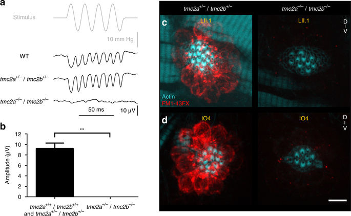
Tmc2a coordinates with Tmc2b to enable mechanotransduction in lateral line hair cells. a Stimulus-evoked microphonic potentials measured from posterior neuromasts with A–P-oriented hair cells from 6-dpf zebrafish larvae. The response in tmc2a −/− /tmc2b −/− is absent, bottom trace. b Graph of mean microphonic potentials from posterior neuromasts with hair cells with A–P orientations (n = 6). **Mann–Whitney test P = 0.0043. c, d Confocal images of hair cells from LII.1 and IO4 neuromasts of tmc2a +/− /tmc2b +/− or tmc2a −/− /tmc2b −/− animals labeled with FM1-43FX (red) and phalloidin (cyan). No dye was observed in LII.1 or IO4 of tmc2a −/− /tmc2b −/− animals. Scale bar = 6 μm