
|
Fig. 2
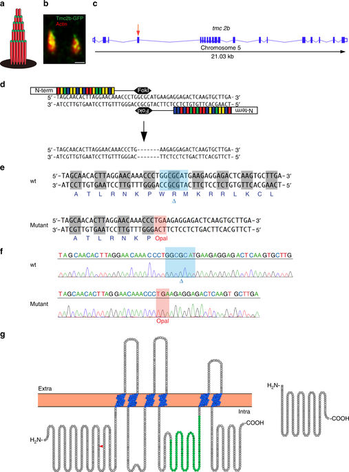
Localization of Tmc2b and TALEN-mediated disruption of the tmc2b gene. a Schematic of the hair bundle. b Tmc2b localizes to the tips of stereocilia in a neuromast. Two lateral line hair cells expressing Tmc2b-GFP (green) demonstrate that the fusion protein localizes to the tips of β-actin-mCherry-labeled stereocilia (red). Scale bar = 0.5 μm. This pattern was observed in 130 of 381 hair cells of somatic transgenics. In transgenic hair cells with weak expression, localization to stereocilia was difficult to recognize. c Graphical representation of the tmc2b genomic locus in zebrafish. Putative exons and splice sites are displayed. Red arrow marks the targeted exon, exon 4. d Segment of exon 4 subjected to genome editing. Two differently engineered TALENs bind their corresponding half-sites to enable FokI dimerization and DNA cleavage. Mutagenesis deleted seven nucleotides. e Amino acid sequences of wild-type and mutant proteins. The alteration results in an opal mutation upstream of all putative transmembrane domains. f Sequencing results of mutagenized and control loci. Blue highlight and blue delta indicates deleted 7-nucleotide stretch absent in mutant. Red highlight denotes the opal mutation that was generated at the site of the TALEN targeting. g (left) Topographical representation of the Tmc2b protein. Red arrowhead indicates point of introduced mutation. Amino acids of putative transmembrane domains are labeled in blue and the TMC domain is in green. (right) Predicted truncated product of mutation