
Image
|
Figure Caption
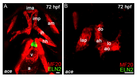
Fig. S3
Analysis of head muscles in ace embryos. (A,B) Confocal Z-stacks of ace mutant embryos double immunostained to detect skeletal muscle (MF20 antibody) and Eln2+OFT cells imagined in the red and green channels, respectively. Representative example of ace mutant embryos containing mispatterned head muscles in pharyngeal arch 1 and 2(n=11/25). Ventral views, anterior up (A). Lateral view, anterior left (B). Please see Figure 1 legend for HM abbreviations. Scale bars=25µm
Acknowledgments
This image is the copyrighted work of the attributed author or publisher, and
ZFIN has permission only to display this image to its users.
Additional permissions should be obtained from the applicable author or publisher of the image.
Full text @ Development