
|
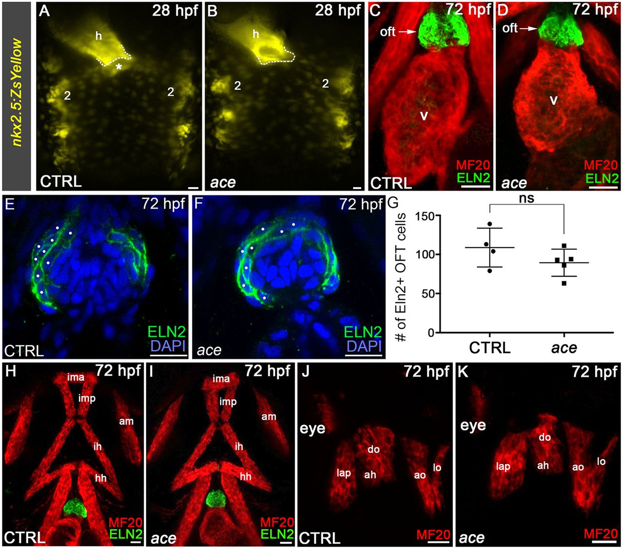
Fig. 6
SHF ventricular and OFT progenitors are uniquely sensitive to perturbations in Fgf8a signaling. (A,B) Compound microscopic images of live 28 hpf control (CTRL) sibling (A) or ace (B, n=33) embryos carrying the nkx2.5:ZsYellow transgene. Dorsal views are shown. Anterior is upwards. The dotted lines demarcate the arterial pole of the heart tube. The asterisk highlights extra-cardiac ZsYellow+ anterior SHF progenitors absent in ace mutants. All 74 control and all ace animals displayed the ZsYellow distributions shown. (C,D) Confocal z-stacks of the ventricle and OFT in 72 hpf CTRL and ace animals double immunostained to detect striated muscle (MF20 antibody) and Eln2+ smooth muscle OFT cells imaged in the red and green channels, respectively. All 76 control and all 25 ace animals contained Eln2+ OFTs. Ventral views. Anterior is upwards. (E,F) Confocal z-stacks of the OFTs in 72 hpf CTRL and ace mutant embryos immunostained to visualize Eln2 (green) and counterstained with DAPI (blue) to detect nuclei. White dots highlight nuclei in cells surrounded by Eln2. (G) Dot plot showing the quantification of Eln2+ OFT cells in 72 hpf control and ace mutant embryos. An unpaired t-test was used to evaluate statistical significance. ns, not significant. (H-K) Confocal z-stacks of pharyngeal regions in 72 hpf control and ace animals double immunostained to detect striated muscle (MF20 antibody) and Eln2+ smooth muscle OFT cells imaged in the red and green channels, respectively. All 76 CTRL and 14/25 ace animals contained the head muscles in the pattern shown. Ventral (H,I) and lateral (J,K) views are shown. Anterior is upwards (H,I) or leftwards (J,K). Number two labels pharyngeal arch 2. h, heart. Ventral pharyngeal arch (PA) 1 (mandibular) muscle: ima, intermandibular anterior. Middle PA1 muscles: imp, intermandibular posterior; am, adductor mandibulae. Dorsal PA1 muscles: lap, levator arcus palatine; do, dilator operculi. Ventral PA2 (hyoid) muscle: ih, interhyal. Middle PA2 muscle: hh, hyohyal. Dorsal PA2 muscles: ah, adductor hyomandibulae; ao, adductor operculi; lo, levator operculi. Cardiac structures: oft, outflow tract; v, ventricle. Scale bars: 25 µm.