
|
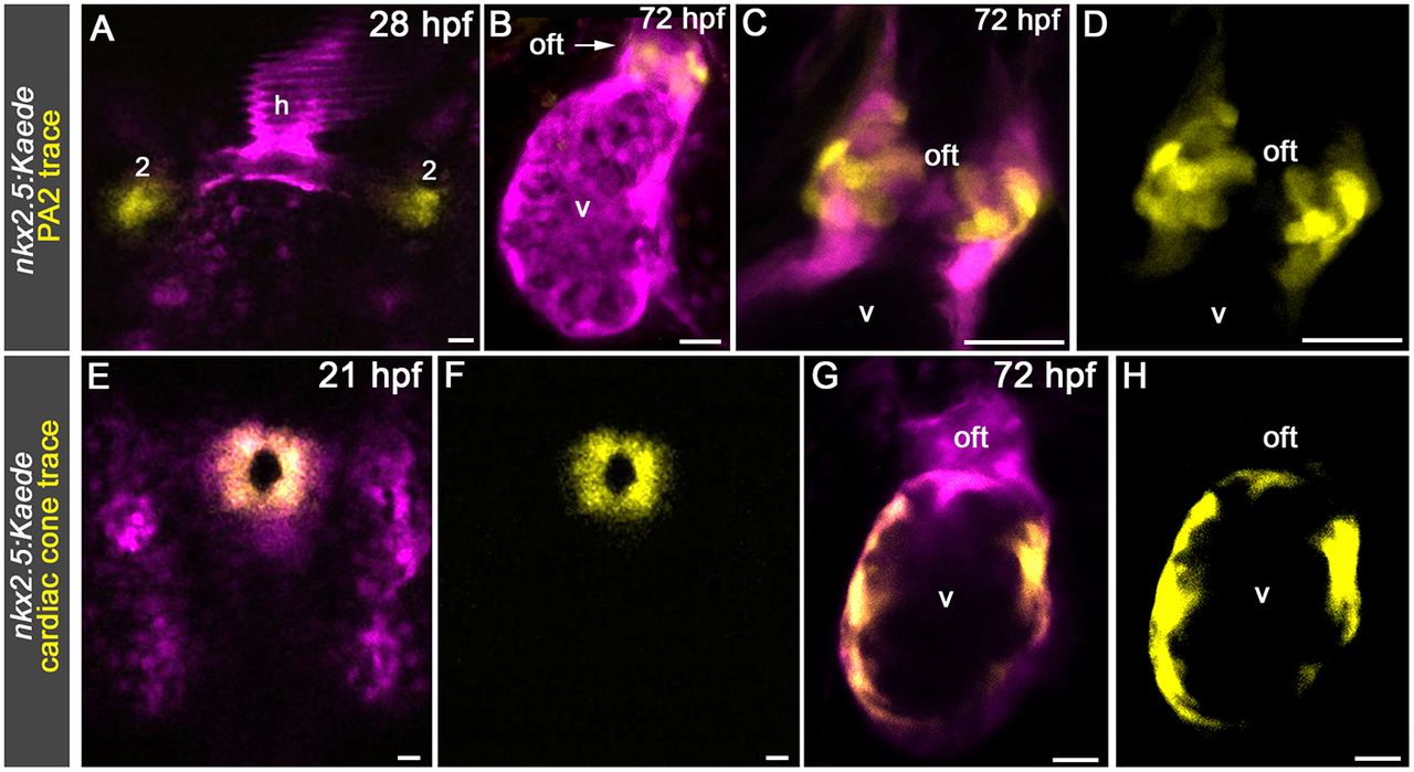
Fig. 4
Unique trajectories of SHF ventricular and OFT progenitors. (A) Confocal z-stack of the pharyngeal region in a 28 hpf Tg(nkx2.5:Kaede) embryo immediately following bilateral photoconversion of Kaede+ cells in pharyngeal arch (PA) 2. A dorsal view is shown. Anterior is upwards. (B) Confocal z-stack of the ventricle and OFT in the same embryo at 72 hpf. The arrow highlights OFT cells carrying the photoconverted Kaede protein. All nine animals contained photoconverted cells in the OFT. (C,D) High-magnification confocal z-stacks of the OFT in a 72 hpf Tg(nkx2.5:Kaede) animal in which Kaede+ cells in PA2 were photoconverted bilaterally at 28 hpf. (E,F) Confocal z-stacks of a 21 hpf Tg(nkx2.5:Kaede) embryo immediately following photoconversion of Kaede+ cells in the cardiac cone. A dorsal view is shown. Anterior is upwards. (G,H) Confocal z-stack of the ventricle and OFT in the same embryo at 72 hpf. All 12 animals contained photoconverted cells in the ventricle but not in the OFT. All animals in A-H were imaged in the green (pseudocolored magenta) and red (pseudocolored yellow) channels. Merged (A-C,E,G) and single-channel (D,F,H) images are shown. Cardiac structures: h, heart; oft, outflow tract; v, ventricle. Scale bars: 25 µm.