
|
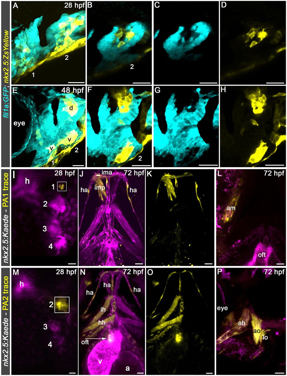
Fig. 2
Visualization and prospective lineage tracing of head muscle and hypobranchial artery endothelial precursors in pharyngeal arches 1 and 2. (A-H) Confocal images of pharyngeal arch (PA) 1 and PA2 in 28 hpf (A-D) and 48 hpf (E-H) Tg(nkx2.5:ZsYellow); Tg(fli1a:GFP) embryos double immunostained for GFP and ZsYellow imaged in the green (pseudocolored cyan) and red (pseudocolored yellow) channels, respectively. Single planes (B-D,F-H) from merged confocal z-stacks (A,E) are shown as merged (B,F) or single-channel (C,D,G,H) images. Left lateral views are shown. Anterior is towards the left. Little to no variability was observed in greater than 30 animals examined. (I,M) Merged confocal z-stacks of pharyngeal regions in live 28 hpf Tg(nkx2.5:Kaede) embryos immediately following unilateral photoconversion of Kaede+ cells (boxed regions) in PA1 (I) or PA2 (M). (J,K,N,O) Confocal z-stacks of pharyngeal regions in the same embryos at 72 hpf shown as merged (J,L,N,P) or single-channel (K,O) images. All five animals with PA1 photoconversion demonstrated tracing to the ipsilateral PA1 head muscles and bilateral HA endothelium. Labeling of the intermandibular anterior muscle crossed the midline in some cases. All 14 animals with PA2 photoconversion demonstrated tracing to the ipsilateral PA2 head muscles, ipsilateral OFT and bilateral HA endothelium. Animals were imaged in the green (pseudocolored magenta) and red (pseudocolored yellow) channels. Dorsal (I,M), ventral (J,K,N,O) and lateral (L,P) views are shown. Anterior is upwards (I-K,M-O) or leftwards (L,P). Numbers label the pharyngeal arches. d, dorsal cluster; v, ventral cluster. Ventral pharyngeal arch (PA) 1 (mandibular) muscle: ima, intermandibular anterior. Middle PA1 muscles: imp, intermandibular posterior; am, adductor mandibulae. Ventral PA2 (hyoid) muscle: ih, interhyal. Middle PA2 muscle: hh, hyohyal. Dorsal PA2 muscles: ah, adductor hyomandibulae; ao, adductor operculi; lo, levator operculi. Vessel: ha, hypobranchial artery. Cardiac structures: oft, outflow tract; a, atrium; v, ventricle. Scale bars: 25 µm.