
|
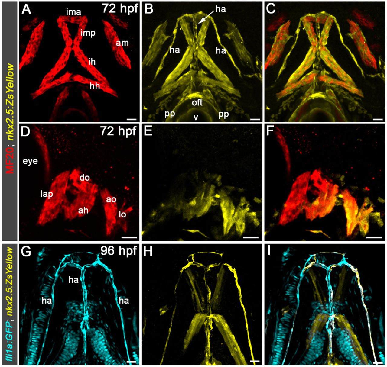
Fig. 1
Identification of ZsYellow+ head muscles and endothelium in Tg(nkx2.5:ZsYellow) larvae. (A-F) Confocal z-stacks of pharyngeal regions in 72 hpf Tg(nkx2.5:ZsYellow) zebrafish larvae double immunostained to detect ZsYellow fluorescent protein and striated muscle (MF20 antibody) imaged in the green (pseudocolored yellow) and red channels, respectively. (G-I) Confocal z-stacks of the pharyngeal region in a 96 hpf Tg(nkx2.5:ZsYellow); Tg(fli1a:GFP) larvae double immunostained to detect ZsYellow and green fluorescent protein (GFP) imaged in the red (pseudocolored yellow) and green (pseudocolored cyan) channels, respectively. Single-channel (A,B,D,E,G,H) and merged (C,F,I) images are shown. Ventral (A-C,G-I) and lateral (D-F) views are shown. Anterior is upwards (A-C,G-I) or leftwards (D-F). For both experiments, little to no variability in the staining patterns was observed in the greater than 30 animals examined. Ventral pharyngeal arch (PA) 1 (mandibular) muscle: ima, intermandibular anterior. Middle PA1 muscles: imp, intermandibular posterior; am, adductor mandibulae. Dorsal PA1 muscles: lap, levator arcus palatine; do, dilator operculi. Ventral PA2 (hyoid) muscle: ih, interhyal. Middle PA2 muscle: hh, hyohyal. Dorsal PA2 muscles: ah, adductor hyomandibulae; ao, adductor operculi; lo, levator operculi. Vessel: ha, hypobranchial artery. Cardiac structures: oft, outflow tract; pp, parietal pericardium; v, ventricle. Scale bars: 25 µm.