
|
Fig. S4
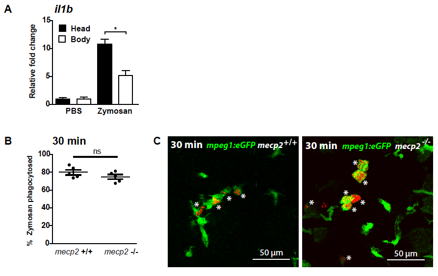
Microinjection of zymosan into the brain of zebrafish larvae (A) Zymosan was injected into the brain of 3 dpf wild type larvae. Quantitative real-time PCR was performed to determine gene expression level of il1b relative to the expression of the housekeeping gene tbp in the dissected heads and bodies of injected larvae. Samples (n=3 with 10 heads or bodies per sample) were taken at 1 hour post injection of zymosan or PBS as a control. The relative fold change of zymosan versus PBS injected samples is shown to account for a possible wounding effect by the injection itself. A One-way ANOVA with Tukey’s post hoc test was used for statistical analysis (*: p<0.05; ns: not significant). (B) The percentage of zymosan particles phagocytes by Tg(mpeg1:eGFP)- positive cells for wild type and mecp2-null larvae using confocal microscopy of samples fixed 30 minutes after injection (n=5 larvae per condition). A Student T-test was used for statistical analysis (ns: not significant). (C) Representative confocal micrographs of a wild type and mecp2-null Tg(mpeg1:eGFP) larvae at 30 minutes post injection. An asterix (*) indicates zymosan phagocytosed by Tg(mpeg1:eGFP)-positive cells.