
|
Fig. 2
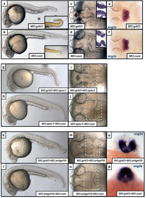
Midbrain-Hindbrain Boundary (MHB) morphogenesis is disrupted following knockdown of grhl3-dependent transcriptional networks. (a,b) Axial length is reduced, neural morphology is disrupted, and fish often exhibit a “curved-tail” phenotype (inset; 96 hpf) in MO:grhl3 injected fish relative to controls. (c,d) The MHB defect in MO:grhl3 injected fish presents as aberrant folding (dorsal view), confirmed by horizontal sections of H&E stained MHB tissue (c,d; inset). (e,f) Although the MHB is mis-folded, MHB patterning markers (such as eng2a) are not differentially regulated. (g–j) Following sub-phenotypic co-knockdown of grhl3 and spec1 (MO:grhl3 + MO:spec1; g,i) the MHB is mis-folded when compared to phenotypically normal MO:spec1 + MO:control injected fish (h,j). (k–p) Following sub-phenotypic co-knockdown of grhl3 and arhgef19 (MO:grhl3 + MO:arhgef19; k), the fish are slightly shorter relative to phenotypically normal MO:arhgef19 + MO:control injected fish (l). Furthermore, the neural tube exhibits an “open” phenotype at the MHB region (dorsal view; m,n); this is highlighted by ISH for the MHB marker eng2a (posterior view of MHB; o). relative to the phenotypically normal MHB seen in MO:grhl3 + MO:control injected fish (p).