
|
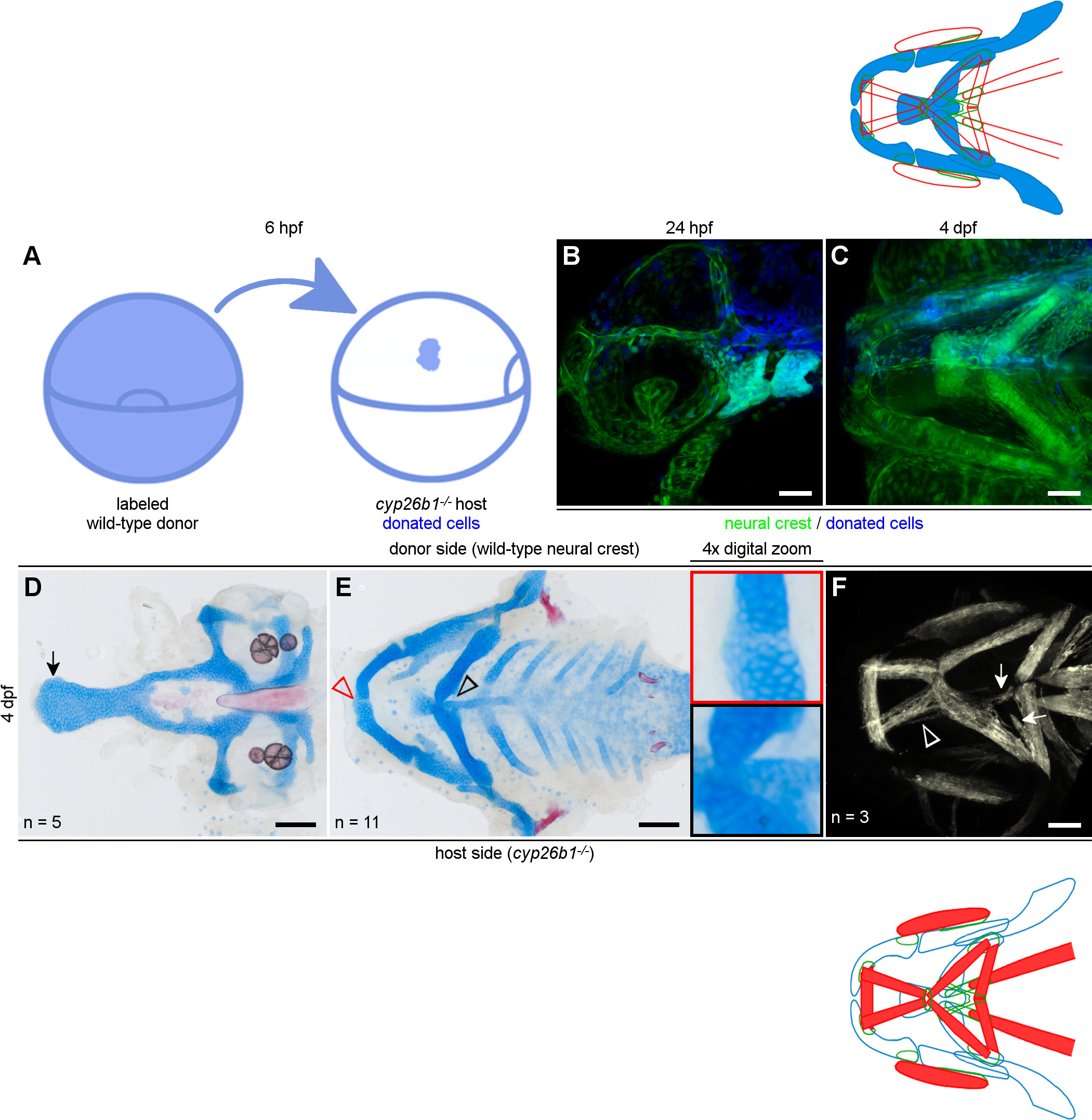
Fig. 4 Cyp26b1 functions in the neural crest to promote craniofacial musculoskeletal patterning. (A) Schematic for transplantation of embryonic cells at 6 hpf. The blue area shown on the right side of A will contribute to the neural crest on one side of the head (B, C). (D-E) Donated neural crest cells improve skeletal phenotypes in a cyp26b1 mutant host. We saw a wider palate with a flared ethmoid plate in mutant hosts (arrow in D). (E) Depletion of Alcian-positive cartilage matrix indicates partial rescue of Meckel’s cartilage fusion (red arrowhead/red inset) and ceratohyal morphogenesis and fusion at the midline is rescued on the donor side (black arrowhead/black inset). (F) On the side of the head without donated neural crest, muscles project ectopically (arrows) and muscle fiber bundles split (arrowhead), compared to normal phenotypes on the donor side. Lateral view in B. All other images ventral view, anterior to the left. Scale bars = 50 μm.