
|
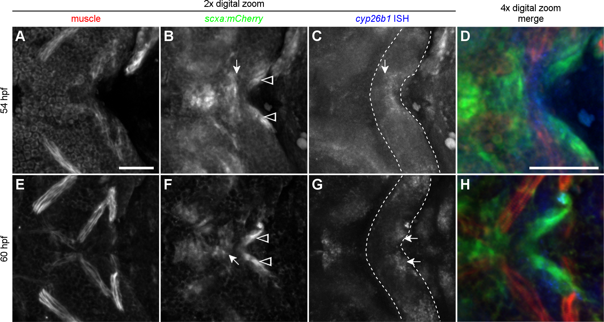
Fig. 9 Cells expressing cyp26b1 separate anterior and posterior tenoblast masses between 54 and 60 hpf. (A-D) 54 hpf zebrafish embryo. (B) Tenoblasts of the future mandibulohyoid junction (arrow) display a flattened posterior boundary and maintain a small distance from tenoblasts lining the posterior edge of the second pharyngeal arch (arrowheads). (C) With the shape of the second arch visible (outline) one can see that cells expressing cyp26b1 line the posterior edge of the arch midline and display a flattened anterior boundary abutting the mandibulohyoid tenoblasts (arrow). (D) With channels combined, cyp26b1 expression (blue, in situ hybridization) labels non-tenoblast cells separating tenoblasts (green, anti-mCherry antibody in scxa:mCherry transgenic) and second arch muscles (red, MF20) that are mostly or entirely cyp26b1-negative. (E-H) 60 hpf zebrafish embryo. (F) Tenoblasts are more compact at the forming mandibulohyoid junction (arrow), and further separated from the sternohyoideus tendon condensations (arrowhead). (G) Strong cyp26b1 expression now labels bilateral cell masses of the posterior edge of the second arch (arrows). (H) Non-tenoblast cells expressing cyp26b1 sit anterior to the sternohyoideus tendon condensations on each side, facing the nearby interhyal muscles. All images ventral view, anterior to the left. Scale bar = 50 μm.