
|
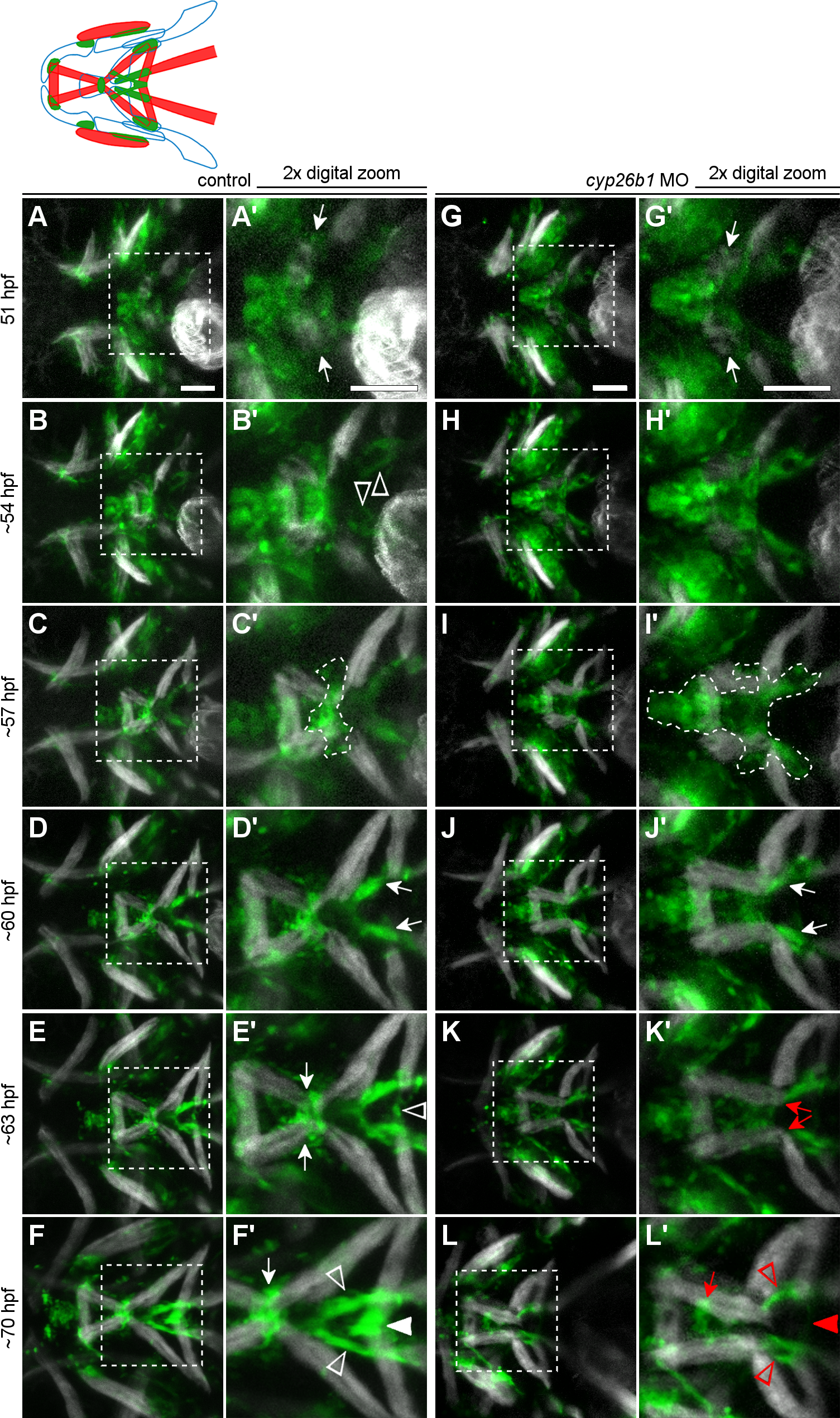
Fig. 7 Loss of Cyp26b1 function disrupts tendon morphogenesis in the second pharyngeal arch midline. (A-L) Stills from S2 Movie. (A,A’) The emerging intermandibularis muscles (arrows) are surrounded by tenoblasts at 51 hpf (B,B’). As tenoblasts converge on the midline, intermandibularis muscles point their posterior tips medially (also see S6B Fig). Meanwhile, tenoblast masses (arrowheads) associated with the hyohyal and sternohyoideus muscles on each side have become visible. (C,C’) By 57 hpf, condensing cells are visible at the mandibulohyoid junction (outline), where scxa-positive cells have segregated from the more anterior group. (D,D’) By 60 hpf, condensation of the sternohyoideus tendons is apparent (arrows). (E,E’) By 63 hpf, tenoblasts attached to each hyohyal muscle connect in the midline and a condensation forms (arrowhead). (F,F’) Approaching 3 dpf, tenoblasts at the mandibulohyoid (arrow) and hyohyal (white arrowhead) junctions are highly condensed between the muscle tips, and sternohyoideus tendons are elongated (open arrowheads). (G,G”) In morpholino-injected embryos, intermandibularis muscles (arrows) emerge normally among a field of tenoblasts around 51 hpf. (H,H’) Tenoblasts migrate slowly toward the midline, and intermandibularis posterior muscles point posteriorly at 54 hpf (also see S6D Fig). (I,I’) Tenoblasts at the mandibulohyoid junction are neither condensing nor separating from adjacent tenoblasts at 57 hpf (outline). (J,J’) Despite surrounding abnormalities, sternohyoideus tendons initiate condensation around 60 hpf (arrows). (K,K’) Connections between intermandibularis posterior muscles and sternohyoideus tendon condensations persist, and at 63 hpf the muscle tips (red arrows) now extend past mandibulohyoid tenoblasts. (L) Compared to controls, the sternohyoideus tendons are poorly condensed and elongated by 70 hpf, and both intermandibularis posterior and both hyohyal muscles connect to either end of these tendon rudiments. (L’) Mesenchymal tenoblasts sit between the intermandibularis posterior muscles (arrow) and no condensation has formed connecting the hyohyal muscles (red arrowhead). All images ventral view, anterior to the left. Scale bar = 50 μm