
|
Fig. 2
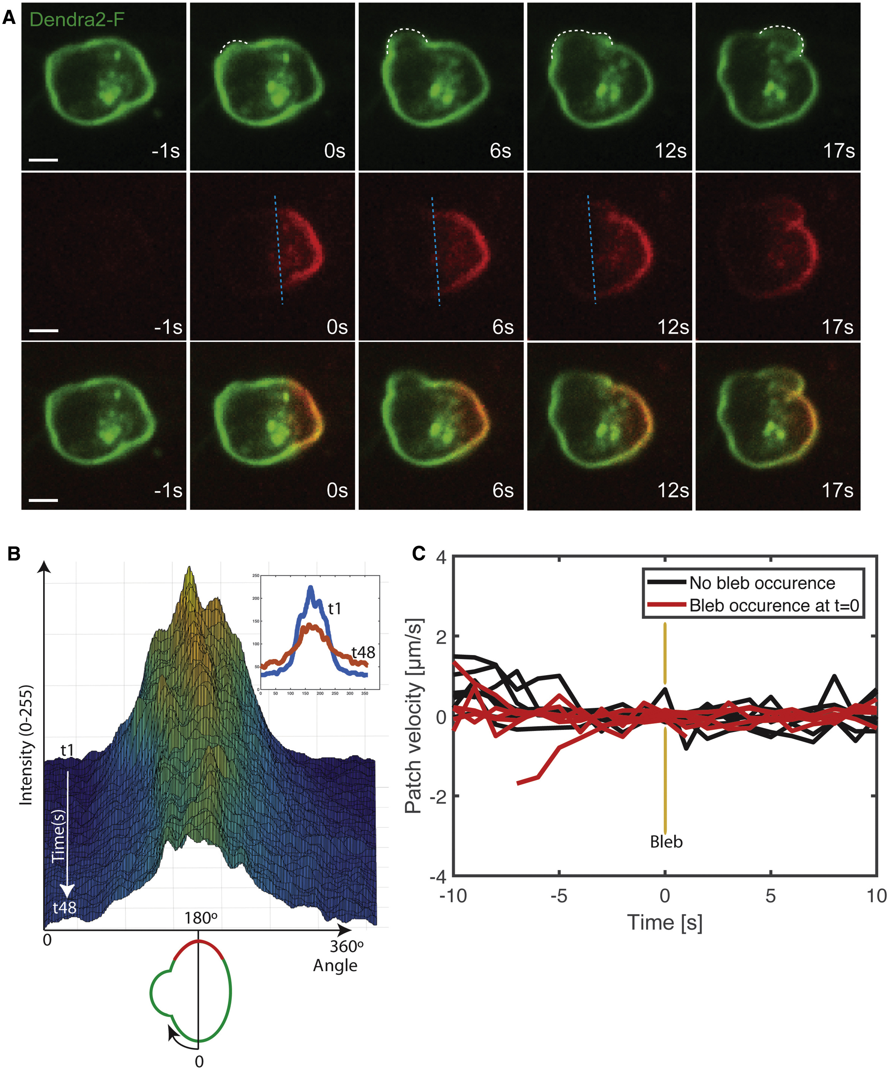
Lack of Membrane Flow in the Direction of the Forming Bleb in PGCs Migrating within Live Embryos
(A) Farnesylated-Dendra2 (Dendra2-F) expressed on the plasma membrane of PGCs from 7 to 8 hr post fertilization (hpf) embryos was photoconverted (green to red) at the border of a forming bleb (white dashed line shows the contours of two sequential blebs, with the blue dashed line marking the edge of the converted area up to the 12 s time point). Cells were imaged at a frequency of one time point per second and the photoconversion time point is presented as zero (0 s). No directional flow with respect to the forming bleb is observed. Seven events of photoconversion next to a forming bleb were analyzed. Scale bar, 5 μm.
(B) 3D surface plot of the converted area showing the intensity change along the circumference (0°–60°) of the cell presented in (A) over 48 s, but no change in the position of the center of the converted area. The inset shows the circumferential intensity in the first (1 s) and the last frame after photoconversion (48 s). A schematic of the cell analyzed is presented below the x axis.
(C) Graph showing the velocity of the center of the photoconverted region in cells where blebs did not form (n = 5, black) and in cells in which bleb inflation occurred next to the photoconverted area (n = 7, red). t = 0 is the time of bleb formation.
See also Figure S1.
Reprinted from Developmental Cell, 43(5), Goudarzi, M., Tarbashevich, K., Mildner, K., Begemann, I., Garcia, J., Paksa, A., Reichman-Fried, M., Mahabaleshwar, H., Blaser, H., Hartwig, J., Zeuschner, D., Galic, M., Bagnat, M., Betz, T., Raz, E., Bleb Expansion in Migrating Cells Depends on Supply of Membrane from Cell Surface Invaginations, 577-587.e5, Copyright (2017) with permission from Elsevier. Full text @ Dev. Cell