
|
Fig. 6
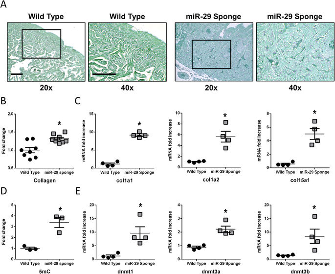
miR-29 family knock-down associates with collagen deposition and DNA methylation increase in the Zebrafish heart. (A) Representative Fast Green Sirius Red staining of Wild Type (left panels) and miR-29-sponge (right panels) Zebrafish ventricles. Collagenous proteins are depicted in light purple and non-collagenous proteins in green. Magnification: 20x in first and third panel and 40x in second and fourth panel. Calibration bar = 25 µm (B) Collagen deposition quantification in sections derived from Wild Type (black circles; n = 8) and miR-29-sponge (gray squares; n = 8) Zebrafish hearts. (C) qRT-PCR analysis of collagen mRNAs in Wild Type (black circles; n = 4) and miR-29-sponge (gray squares; n = 4) Zebrafish hearts expressed as fold increase versus Wild Type samples. (D) Global DNA methylation quantification of 5mC in Wild Type (black circles; n = 3) and miR-29-sponge (gray squares; n = 3) Zebrafish hearts expressed as fold-change versus Wild Type samples. (E) qRT-PCR analysis of dnmt mRNAs in Wild Type (black circles; n = 4) and miR-29-sponge (gray squares; n = 4) Zebrafish hearts expressed as fold increase versus Wild Type samples. *p < 0.05 Vs Wild Type.