
|
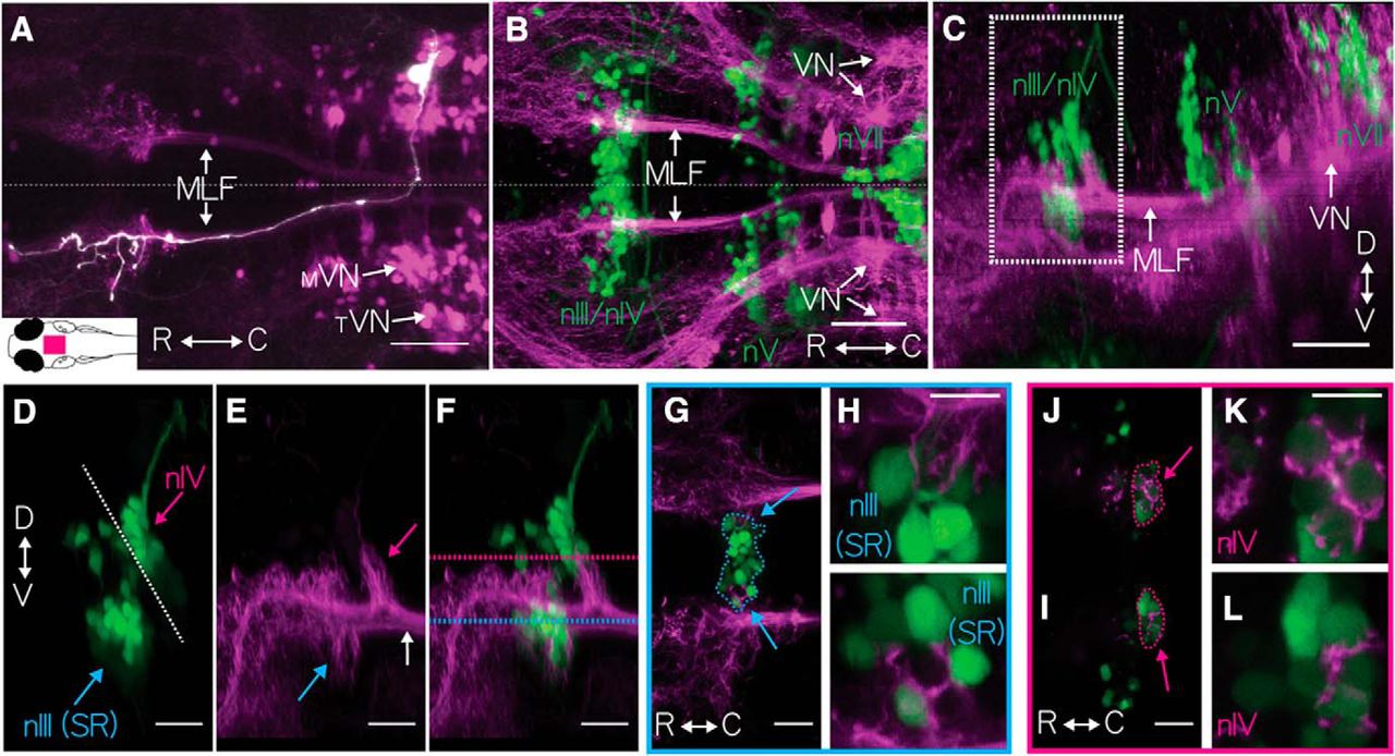
Fig. 1
Vestibular nucleus neurons labeled in Tg(−6.7FRhcrtR:gal4VP16). A, The expression pattern of Tg(−6.7FRhcrtR:gal4VP16); Tg(UAS-E1b:Kaede)s1999t (purple) is shown as a horizontal MIP, with one vestibular neuron, colabeled by focal electroporation of gap43-EGFP (white). Arrows point to the tangential (TVN) and medial vestibular nuclei (MVN) and the MLF. Inset, Schematic of a dorsal view of a larval zebrafish. Magenta rectangle represents the location of the image. Scale bar, 50 μm. Horizontal (B) and sagittal (C) MIP of vestibular neurons in Tg(−6.7FRhcrtR:gal4VP16);Tg(UAS-KillerRed) (purple);Tg(isl1:GFP) (green, image γ = 0.5) showing cranial motoneuron somata from nIII/nIV, nV, and nVII (green text). Arrows indicate neurons in the vestibular nuclei (VN) and the MLF. Scale bar, 50 μm. D–F, Close-up of white boxed region in C, showing major branch patterns of vestibular neuron axon fascicle (purple) relative to extraocular motoneurons (green). D, Motoneurons from Tg(isl1:GFP) (green) in nIV (magenta arrow), superior rectus motoneurons of nIII (cyan arrow), and the midbrain/hindbrain boundary (white dotted line). E, Branches of the vestibular neuron axon fascicle (purple), emerging from the MLF (white arrow) in Tg(−6.7FRhcrtR:gal4VP16);Tg(UAS-KillerRed), projecting to nIV (magenta arrow) and nIII (cyan arrow). F, Merge of D and E. Scale bar, 20 μm. G–I, Broad and close-up views of vestibular neuron axonal projection (purple) to nIII cell bodies (green), taken at the horizontal plane delineated by the cyan dotted line in F, SR motoneurons (nIII) encircled in cyan. G, Cyan arrows localize close-ups in H and I. Scale bar, 10 μm. J–L, Broad and close-up view of vestibular neuron axonal projection (purple) to nIV cell bodies (green), taken at the horizontal plane delineated by the magenta dotted line in F, SO motoneurons (nIV, green) encircled in magenta. J, Arrows point to close-up in K and L. Scale bar, 10 μm.