
|
Fig. 6
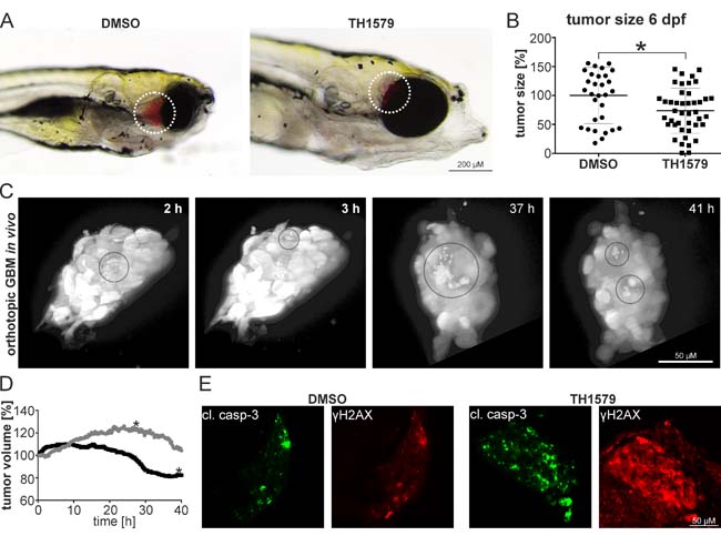
MTH1 inhibitors target GBM and GBM stem cells in vivo. GBM #18-CMV-LUC cell enriched for the CD133+ population have been orthotopically injected into zebrafish embryos. 6 days post injection, embryos exposed to 50 µM TH1579 displayed smaller tumors A. Quantification by luminescence measurements in single embryos showed 26.4 % smaller tumors in TH1579 treated embryos (n = 43) compared to DMSO controls (n = 31; p = 0.011) B. Still images of real-time light sheet microscopy on orthotopic xenotransplants exposed to 50 µM TH1579 apoptotic cells encircled C. Determination of tumor volume of xenotransplant. Grey circle: DMSO control, black circle TH1579 treated (tumor shown in (C). The asterisks mark the time-point when the transplants started leaving the focal plane D. Immunocytochemistry on cleaved caspase as well as y-H2AX in orthotopic xenotransplants treated for 5 days with 50 µM TH1579 or DMSO E.