
|
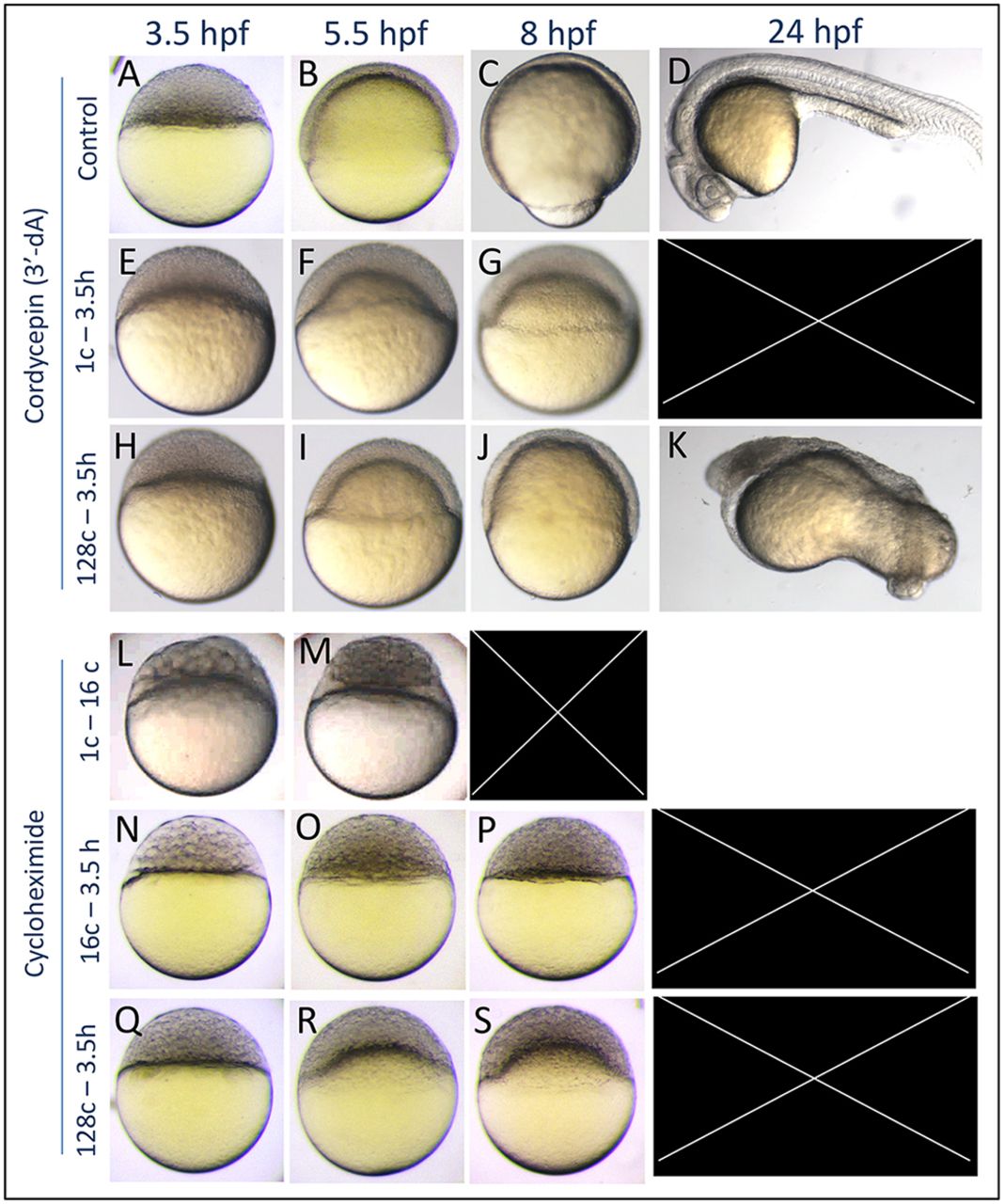
Fig. 3 Epiboly defects caused by 3′dA and CHX treatments. (A-D) Untreated control embryos. (E-G) Embryos that were treated with 3′-dA from the one-cell stage to 3.5 hpf undergo developmental arrest and cytolysis before 24 hpf (n=40). (H-K) 3′-dA treatment from the 128-cell stage to 3.5 hpf caused a delay of epiboly and gross patterning defects (n=30). (L,M) Translation inhibition by CHX treatment that was initiated at the one-cell stage affected early development and caused early lethality (n=20), whereas CHX treatment initiated at the 16-cell stage (N-P) resulted in developmental arrest at the oblong stage (n=61). Notice the larger cells at 3.5 hpf, suggesting defects in cell division. (Q-S) Treatment that was initiated at the 128-cell stage resulted in epiboly arrest followed by mortality (n=66).