
Image
|
Figure Caption
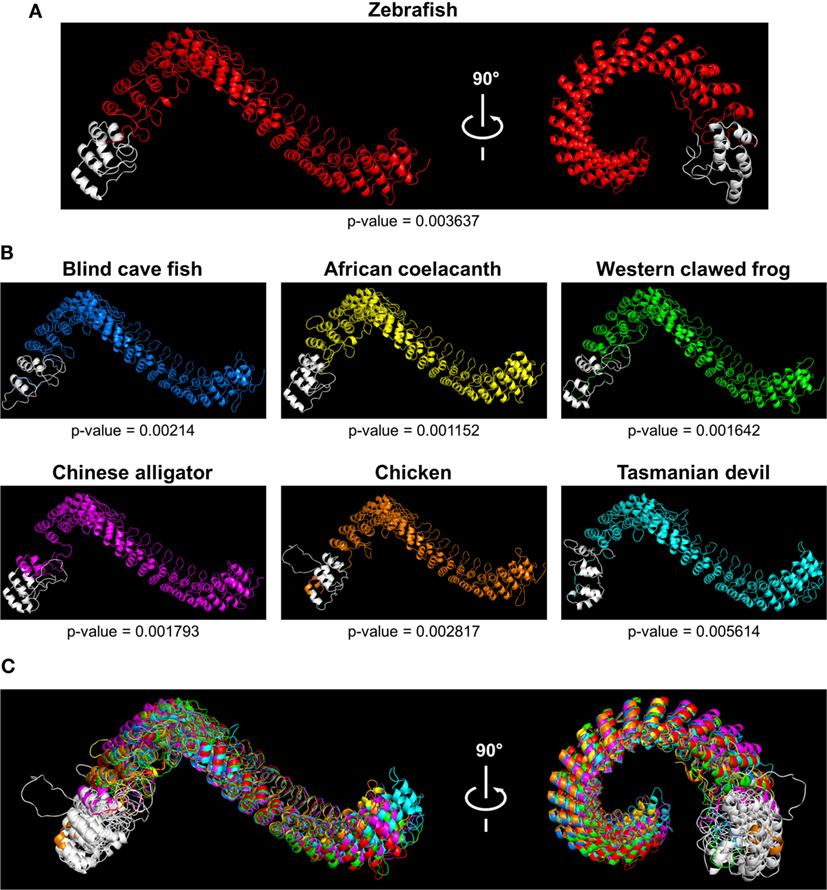
Fig. 2
Tridimensional models of Caiap structures in different species. (A,B) 3D models showing Caiap proteins from zebrafish (Danio rerio) (A), and blind cave fish (Astyanax fasciatus mexicanus), western clawed frog (Xenopus tropicalis), African coelacanth (Latimeria chalumnae), Chinese alligator (Alligator sinensis), chicken (Gallus gallus), and Tasmanian devil (Sarcophilus harrisii) (B) with corresponding accuracies. (C) The 3D Caiap models from all species shown in (A,B) were superimposed. The CARD domains are shown in white.
Acknowledgments
This image is the copyrighted work of the attributed author or publisher, and
ZFIN has permission only to display this image to its users.
Additional permissions should be obtained from the applicable author or publisher of the image.
Full text @ Front Immunol