
|
Fig. s4
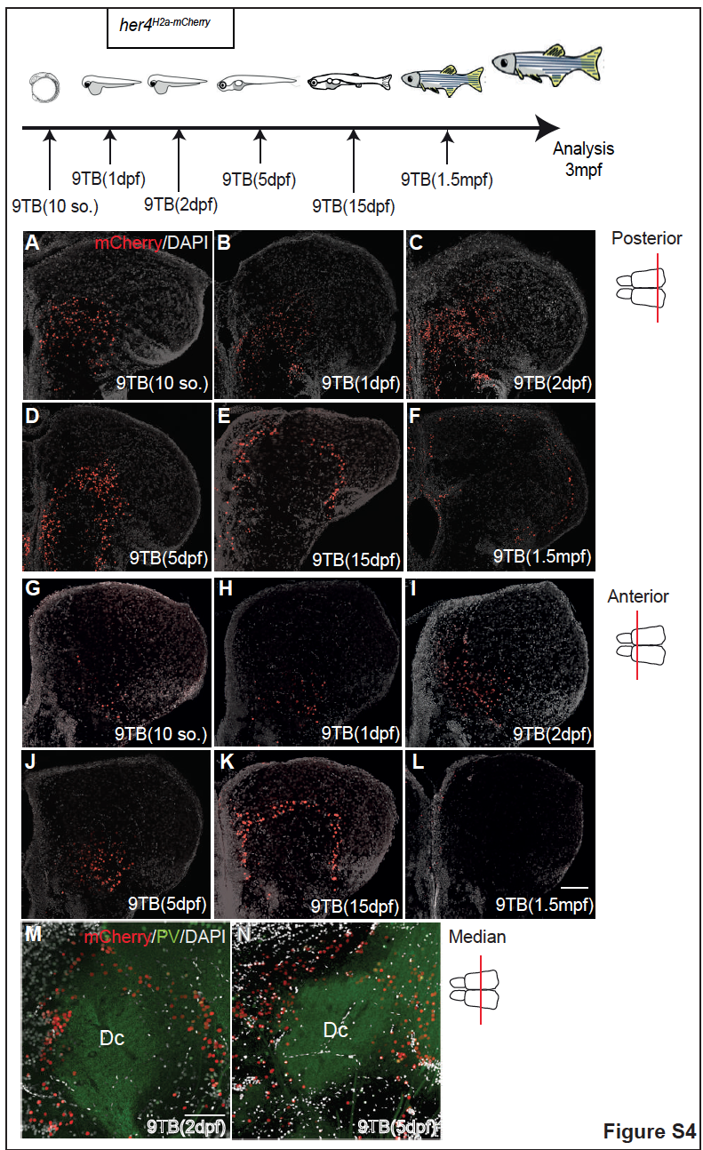
Zebrafish pallial neurogenesis follows a sequential stacking process : anterior and posterior analysis, related to Fig.2. A-L. Localization at posterior and anterior adult pallial levels of the mCherry-positive neurons born from her4-positive RG. Position of mCherry-positive neurons on posterior (A-F) and anterior (G-L) 3mpf her4H2a-mCherry adult pallium cross sections observed in confocal microscopy (with DAPI counterstain) following 9TB induction at the stages indicated. M, N. Localization, at median pallial levels, of the neurons born at 2 and 5dpf relative to Dc. High magnification views of the confocal images shown in Fig.2C’ and D’, respectively, stained for mCherry and PV and counterstained with DAPI. Scale bars: 50µm.