
|
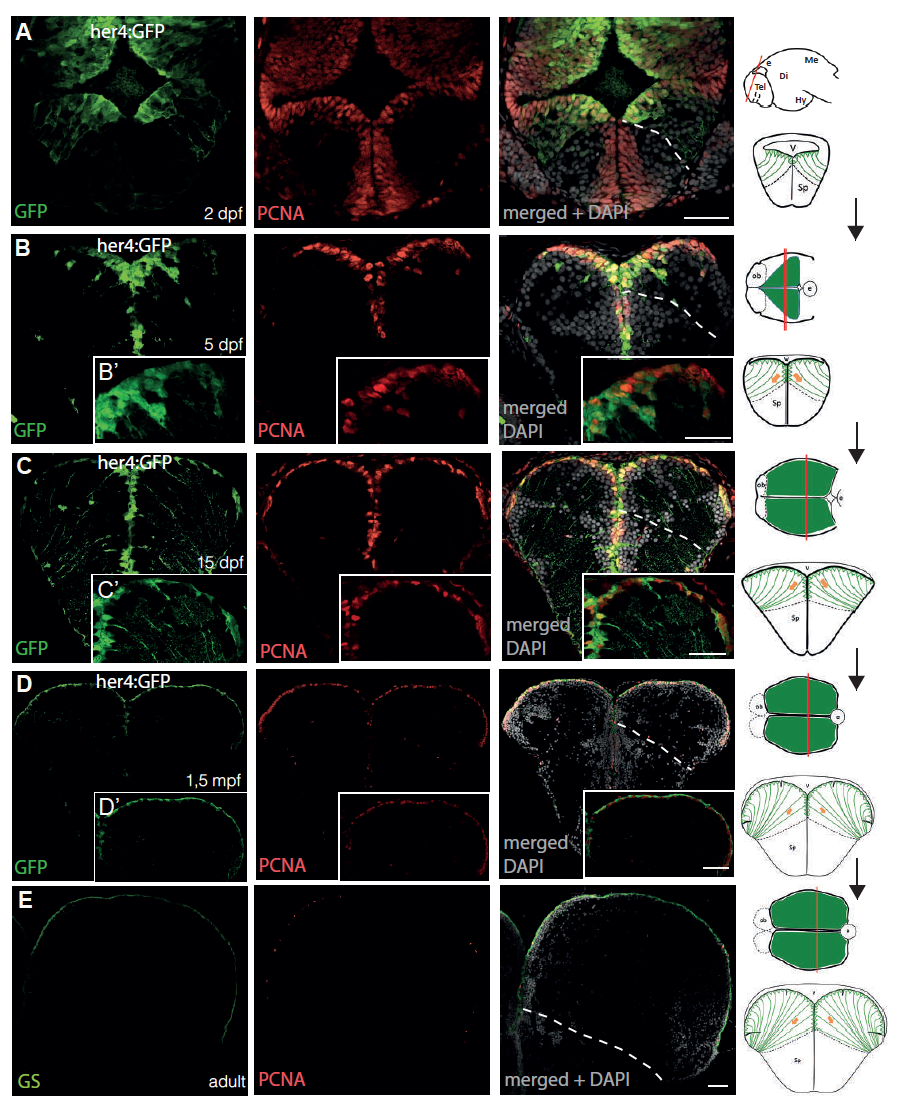
Fig. s2
Continuous and direct neurogenesis from radial glial (RG) cells in the zebrafish pallium from embryo to adulthood, related to Fig.1. A-E. Cross sections of transgenic her4:GFP telencephali at 2dpf (A), 5dpf (B), 15dpf (C), 1.5mpf (D) and adult stage (E) at medial antero-posterior levels (see schemes on the right), immunostained for GFP or the RG marker Glutamine Synthase (green, left panel, as indicated), the proliferation marker PCNA (red, middle panel) and counterstained with DAPI (right panel). High magnifications in insets. White dashed line: position of the pallium/subpallium boudary. Scale bars: A, B inset, C inset : 20μm; D inset, E: 50μm. Abbreviations: Di: diencephalon; e: epiphysis; Hy: hypothalamus; Me: mesencephalon; ob: olfactory bulb; Sp: subpallium; Tel: telencephalon, V: ventricle.