
|
Fig. 9
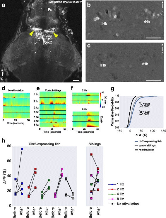
Effect of optogenetic stimulation of the thalamus on habenula activity. a Expression of ChR2-eYFP in the thalamus (arrowheads) of a 5-day-old GAL4s1020t, UAS:ChR2-eYFP, elavl3:GCaMP6f fish. b, c Activity in the habenula of a ChR2-expressing fish, with (b) and without (c) blue LED stimulation of the thalamus. The images show the maximum projections of F/F0 images for a 25-second period after blue LED illumination, following subtraction of maximum projections of the period before illumination (i.e., difference in activity before and after stimulation). d–f Heatmaps showing temporal activity from habenula neurons segmented in fish with (e, f) and without (d) ChR2. In panels e (n = 3 fish) and f (n = 2 fish), a blue light pulse was given at the time indicated by the black dashed line and at the specified frequency. g Cumulative distribution of mean response amplitude, 10 seconds after stimulation in ChR2-expressing and control fish and a randomly chosen 10 second period in fish with no stimulation. All stimulation frequencies were combined. The fish with ChR2 showed increased ∆F/F0 after optogenetic stimulation. Test statistic and P values were obtained using the Kolmogorov–Smirnov test. The gray * (bottom) is the result of comparison between control siblings and Chr2-expressing fish, while the black * (top) is the result of comparison between no stimulation and Chr2-expressing fish. h Mean amplitude before and after optogenetic stimulation at different frequencies. Each square stands for a stimulus trial. Scale bar = 25 μm. Pa pallium, a anterior, p posterior, lHb left habenula, rHb right habenula