
|
Fig. 4
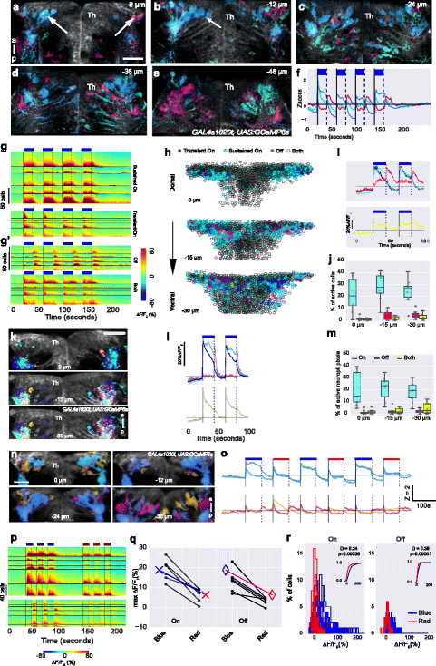
The response of thalamic neurons to irradiance change. a–e Activity in five different focal planes of a 5-day-old fish expressing GCaMP6s in thalamic neurons (arrows). Numbers indicate depth. The colors represent K-means cluster centers shown in panel f, with blue indicating ON responses and magenta indicating OFF responses; cyan pixels have a response to both light ON and OFF. g–j Quantitative analysis of response of anterior thalamic neurons of GAL4s1020t, UAS:GCaMP6s fish (5–6 dpf) to pulses of blue light. Note that this driver is not expressed in afferent retinal ganglion cells. g Heatmaps of individual cells in five example fish, showing major classes of responses seen in thalamic neurons: sustained or transient excitation to light ON, light OFF, or both ON and OFF (yellow). g and g’ have different scales. h Responses in cells from 10 fish at three different focal planes. Four pulses of blue light were given and imaging was done at 7 Hz. Segmented cells in all fish overlaid and colored by their response. i Traces showing mean responses of cells in panel h for two blue pulses. j Percentage of cells responding to light ON, OFF, or both ON and OFF. k Neuropil responses to pulses of light. Pixels with different response classes from all fish were pseudo-colored and overlaid on an average image from a 5-dpf, GAL4s1020t, UAS:GCaMP6s fish. l Average traces of responses in panel k. m Percentage of neuropil pixels responding to light ON, OFF, or both ON and OFF. n–r Thalamus response to blue and red light. n Spatial distribution of responses, color coded according to the K-means cluster centers in panel o, with blue pixels showing a sustained response to light ON, while magenta pixels and orange pixels are a mixture of responses to both ON and OFF. Z is the Z-score. p Heatmaps of cells responding to three pulses of blue light followed by three pulses of red light in n = 6 GAL4s1020t, UAS:GCaMP6s fish. Cells were classified as responding to light ON or OFF. While the same cells responded to both blue and red light, the amplitude of responses were lower to red light. q Peak amplitude of response during light ON and OFF is higher to blue light than red light. Each circle represents one fish and lines join data points from the same fish. Crosses and diamonds represent median amplitude. r Histogram showing amplitude of responses during blue (blue traces) and red (red traces) light ON (left panel) and OFF (right panel). Each trace is response distribution from all cells in a single fish. P values and test statistic (D) were obtained using Kolmogorov–Smirnov test on cumulative response distribution from all fish shown in the inset in r. Th thalamus