
|
Fig. 2
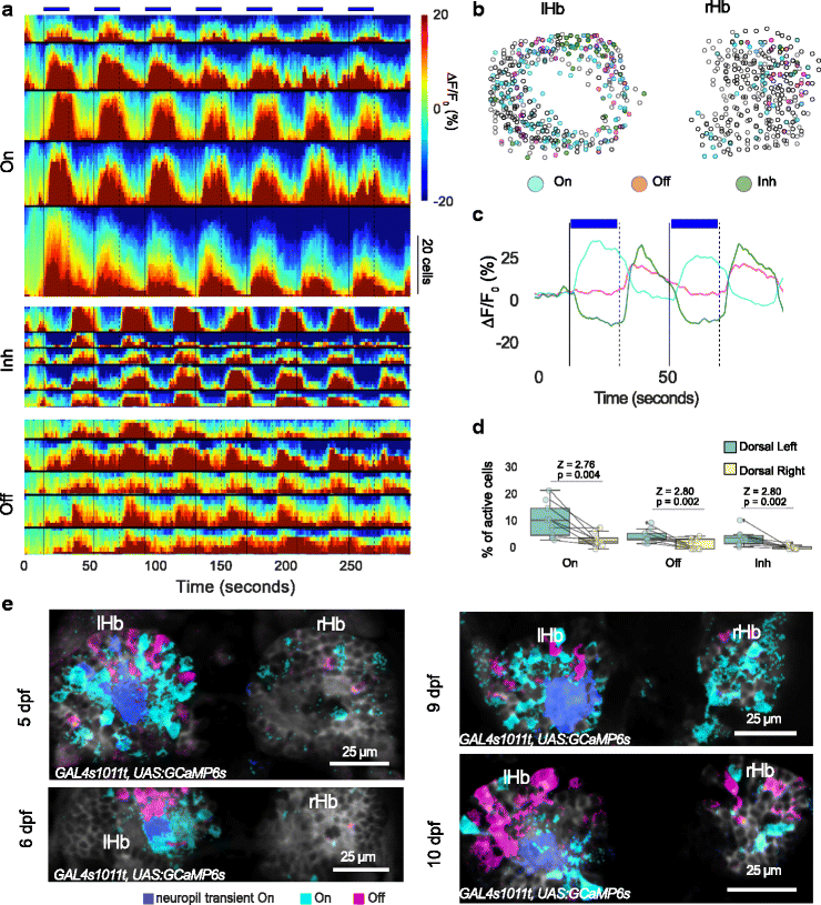
Response of habenula neurons to pulses of light. a–d The dorsal habenula response to 7 pulses of blue light in 10 fish (GAL4s1011t, UAS:GCaMP6s, 5–7 dpf). a Heatmaps from 5 example fish showing responses in cells that were classified as ON, OFF, or Inhibitory (Inh). The colors indicate ∆F/F0, as shown in the color bar. Responses in each fish are sorted in ascending order of mean ∆F/F0. Black horizontal lines separate each fish. The bold vertical lines correspond to light onset while the dashed lines indicate offset. The presence of light is also indicated by the blue bars. The height of the heatmaps represents the number of cells as indicated by the vertical scale bar. b Overlay of cells segmented from all fish. A small circle was drawn around the centroid of the segmented cell. Three main classes of activity are shown. Cyan indicates cells responding to light ON (ON), green cells are inhibited by light (Inh), and magenta cells are activated in the absence of light (OFF). Hollow circles did not show an evoked response. The gap in the left habenula indicates the neuropil region. c Averaged traces from the cells in panel b, showing the response of different classes for the first two pulses of light. d Boxplots showing distribution of cells responding to different classes in the left and right habenula. Each circle is one fish and the line joins data points from the same fish. P values and test statistic (Z) were obtained using Wilcoxon signed rank test. e K-means clustering of pixels in the habenula of fish from 5 to 10 dpf as indicated. Pixels are colored blue if they respond to light ON and magenta if they respond to light OFF. Data from each fish was analyzed separately. Individual traces of the cluster centroids are not shown here but are similar to Fig. 1f. All fish have a response to light onset in the dorsal left neuropil. Anterior is to the top in all panels. rHb right habenula, lHb left habenula