
|
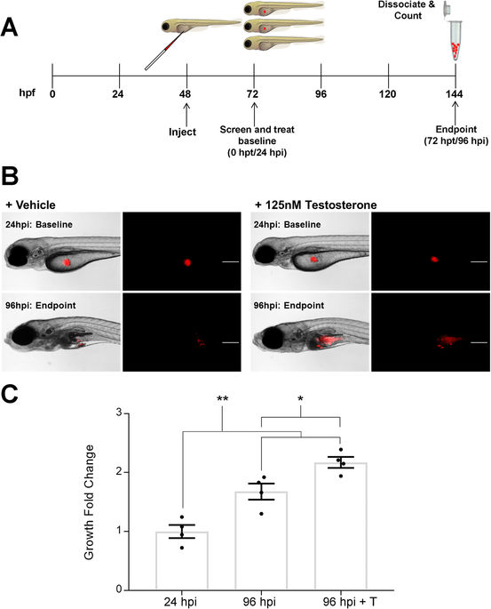
Fig. 1
Xenografted LNCaP cells proliferate significantly in vivo with the addition of testosterone. (A) Schematic of in vivo zebrafish XT microinjection and cell proliferation assay. (B) Representative brightfield and fluorescent images of casper embryos injected with CMTMR labeled LNCaP cells at 24 hpi (baseline) and 96 hpi (endpoint) without and with the addition of testosterone. Scale bar = 200 microns. (C) Quantification of XT LNCaP cell engraftment and fold change ex vivo without and with the addition of 125 nM testosterone (T). LNCaP cells engrafted and proliferated in the XT model indicated by the significant increase in the fold change from baseline numbers (24 hpi) to the endpoint (96 hpi), which increased even more with the addition of testosterone. Error Bars = Mean ± SEM (N = 4); *P < 0.05, **P < 0.01 for significant increase in number of cells determined using the Student’s t-test. Groups of 20 embryos were sacrificed per replicate.