
Image
|
Figure Caption
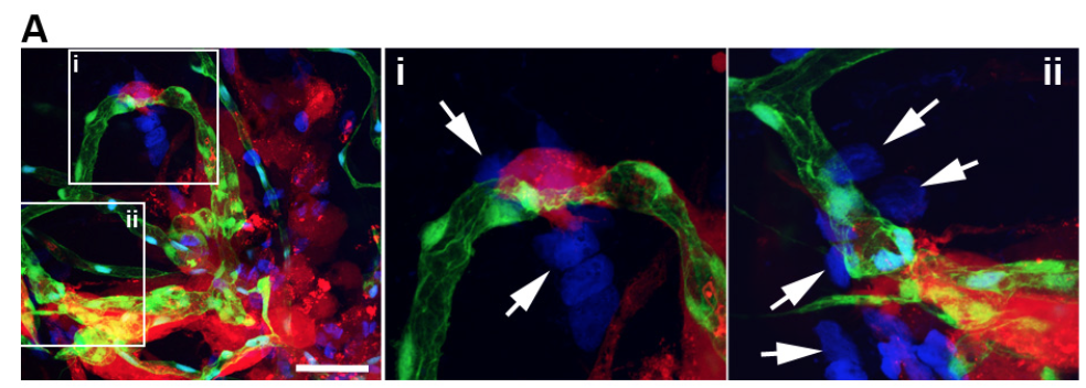
Fig. s3
U87-RFP tumor cells were injected into the flk1: EGFP zebrafish brain as described in method section. DAPI was injected into the blood circulation though caudal vein 1 hour before the confocal imaging of the zebrafish brain. Two areas (i and ii) are magnified right, arrows indicate the DAPI leakage, scale bar indicates 100μm。
Acknowledgments
This image is the copyrighted work of the attributed author or publisher, and
ZFIN has permission only to display this image to its users.
Additional permissions should be obtained from the applicable author or publisher of the image.
Full text @ Sci. Rep.