
|
Fig. 6
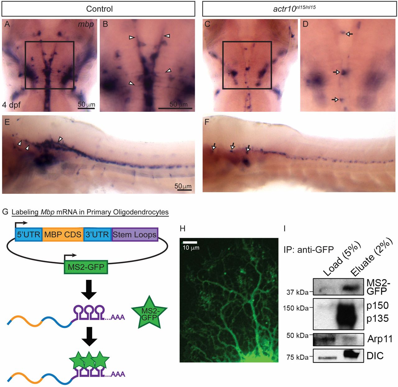
Dynein and dynactin are associated with Mbp mRNA granules. (A) ISH shows robust levels of mbp mRNA in a representative control larva at 4 dpf, and higher magnification (B) shows mbp processes in the control (arrowheads) (n = 47/47). In contrast, an actr10nl15/nl15 mutant (C) has a punctate mbp phenotype, and higher magnification of the same image (D) shows mbp-positive cell bodies (arrows) but no processes (n = 18/18). (E) A lateral view of the brain and spinal cord of a WT animal shows mbp mRNA in processes (arrowheads), while actr10nl15/nl15 mutant animals (F) have cell bodies (arrows) but reduced mbp mRNA-bearing processes. (G) A bidirectional construct expressing MS2-GFP, a RNA-binding reporter, as well as MBP 5′UTR, CDS, and 3′UTR tagged with MS2 stem loops was electroporated into purified rat oligodendrocytes. MS2-GFP binds to stem loops to allow visualization of Mbp mRNA motility. (H) A primary rat oligodendrocyte expressing MS2-GFP–labeled Mbp mRNA imaged using spinning-disk confocal microscopy shows distribution of Mbp mRNA throughout the oligodendrocyte processes. (I) Lysates from primary rat oligodendrocytes expressing the MS2-GFP–labeled Mbp mRNA construct were immunoprecipitated using an anti-GFP antibody and probed with p150Glued, DIC (dynein intermediate chain), and Actr10/Arp11 antibodies (n = 4 independent experiments).