
|
Fig. s3
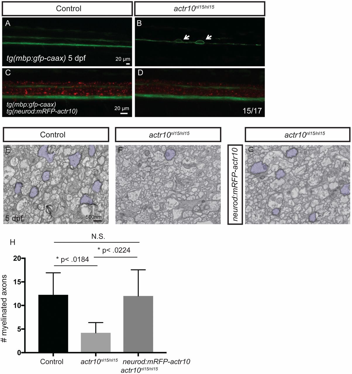
Stable expression of actr10 in neurons suppresses myelination defects in actr10nl15/nl15 mutants (related to Fig. 5). (A–D) Fluorescent micrographs showing tg(mbp:gfp-caax) and tg(neurod:mRFP-actr10) expression in zebrafish of the indicated genotypes harboring the indicated transgenes at 5 dpf. (A) The ventral and dorsal spinal cords are normal in a WT control animal lacking the neurod:mRFP-actr10 transgene. (B) Large caliber swellings (arrows) are present in an actr10nl15/nl15 mutant lacking the neurod:mRFP-actr10 transgene. (C and D) Swellings are not observed in a WT control (C) and are ameliorated in actr10nl15/nl15 mutants harboring the neurod:mRFP-actr10 transgene (n = 15/17). (E–G) TEM images of the dorsal spinal cord show myelinated axons (pseudocolored purple) in WT control animals (E) (n = 4), actr10nl15/nl15 mutants lacking the stable neurod:mRFP-actr10 transgene (F) (n = 3), and actr10nl15/nl15 mutants harboring the stable neurod:mRFP-actr10 transgene (G) (n = 4). (H) Quantification shows that stable neuronal expression of actr10 suppresses actr10nl15/nl15 mutant myelination defects to WT levels (P < 0.99, N.S.), while there is a significant difference between control and actr10nl15/nl15 mutants (*P < 0.018) and actr10nl15/nl15 mutants and actr10nl15/nl15 mutants with the neurod:mRFP-actr10 transgene (*P < 0.022).