
|
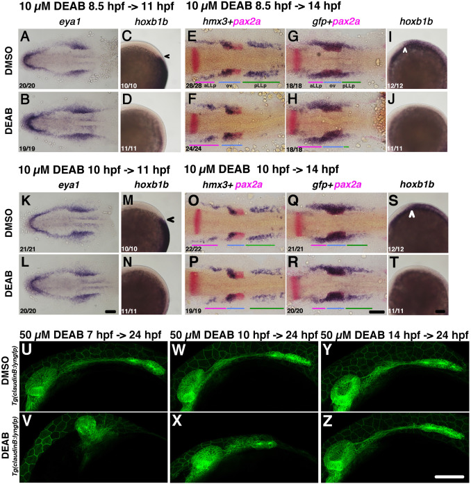
Fig. 5
Removal of RA activity before 10 hpf suppresses pLLp formation but does not affect aLLp formation. (A-T) Duration of the inhibitor treatments and timing for fixation are indicated on the top-left of a group of pictures. After drug treatment embryos were immediately fixed and subjected to in situ hybridization. Drugs for treatment (0.5% DMSO or 10 µM DEAB) are shown on the left. Numbers in the bottom-left indicates numbers of embryos showing the phenotype. (A, B, K, L) Flat-mounted embryos at 11 hpf stained with eya1. Anterior to the left. (E-H, O-R) Flat-mounted embryos at 14 hpf stained with hmx3+pax2a (E, F, O, P) and gfp+pax2a (G, H, Q, R). Anterior to the left. (C, D, I, J, M, N, S, T) Lateral views of 11 hpf (C, D, M, N) and 14 hpf (I, J, S, T) embryos stained with hoxb1b. Anterior is oriented to the top for C, D, M, N, whilst anterior to the left for I, J, S, T. Black and white arrowheads indicate the anterior edge of the neural hoxb1b expression. (U-Z) Longer incubation with DEAB until 24 hpf. Durations for incubation are indicated on top of a pair of pictures in the same column. Drugs treated (0.5% of DMSO and 50 µM of DEAB) are on the left side. Colored bars indicate the positions of the aLLp, otic vesicle and pLLp. Abbreviations: aLLp: anterior lateral line placode, ov: otic vesicle, pLLp: posterior lateral line placode. Scale bars: 100 µm.
Reprinted from Developmental Biology, 431(2), Nikaido, M., Acedo, J.N., Hatta, K., Piotrowski, T., Retinoic acid is required and Fgf, Wnt, and Bmp signaling inhibit posterior lateral line placode induction in zebrafish, 215-225, Copyright (2017) with permission from Elsevier. Full text @ Dev. Biol.