
|
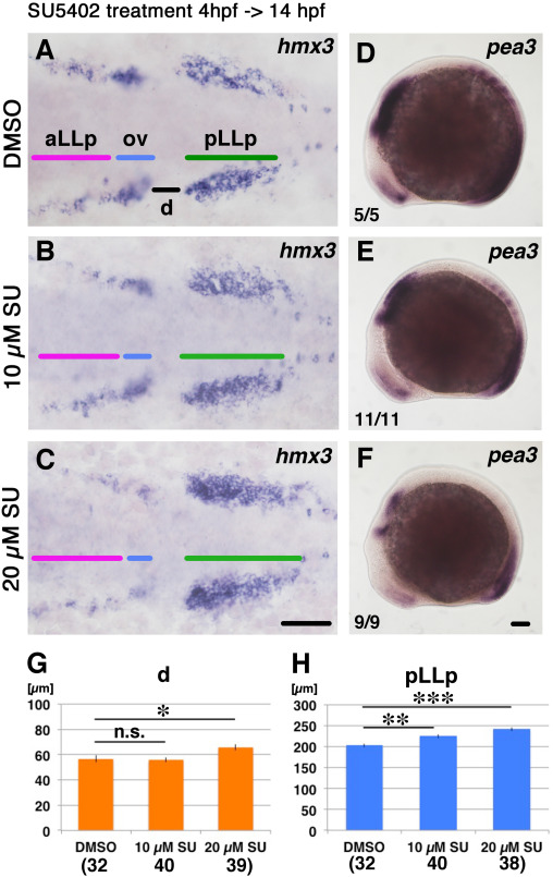
Fig. 4
The aLLp is reduced and not fused to the pLLp after loss of Fgf signaling. Duration of the inhibitor treatment and timing for fixation for A-F are indicated on the top-left. After drug treatment embryos were fixed and subjected to in situ hybridization. Drugs for treatment (0.5% DMSO or SU5402) are shown on the left. Numbers in the bottom-left of D-F indicate numbers of embryos showing the phenotype. (A-C) Flat-mounted embryos at 14 hpf stained with hmx3. Anterior to the left. (D-F) Left-side views of 14 hpf embryos stained with pea3. Anterior to the left. Scale bars: 100 µm. (G, H) Measurement of lengths indicated in panel (A), d and pLLp. Concentration of drug and numbers of hmx3 expression domains examined are shown below each bar. *: p<0.05, **: p<0.01, ***: p<0.001. n.s. is “not significant”. Colored bars indicate the positions of the aLLp, otic vesicle and pLLp. Abbreviations: aLLp: anterior lateral line placode, ov: otic vesicle, pLLp: posterior lateral line placode.
Reprinted from Developmental Biology, 431(2), Nikaido, M., Acedo, J.N., Hatta, K., Piotrowski, T., Retinoic acid is required and Fgf, Wnt, and Bmp signaling inhibit posterior lateral line placode induction in zebrafish, 215-225, Copyright (2017) with permission from Elsevier. Full text @ Dev. Biol.