
|
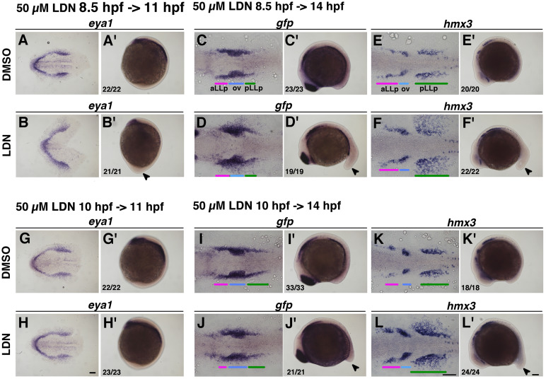
Fig. 2
Removal of Bmp activity leads to expansion of the pLLp. Duration of the inhibitor treatment is indicated on the top-left for pictures. The concentration of DMSO was 0.5% and 50 µM of LDN193189. After drug treatment embryos were immediately fixed and subjected to in situ hybridization. Numbers in the bottom-left indicate number of embryos showing the phenotype out of all examined embryos. (A-F, A’-F’) Inhibitor treatment started at 8.5 hpf (80% epiboly) stages. (G-L, G’-L’) Inhibitor treatment started at 10 hpf stages. (A, B, G, H) Flat-mounted embryos at 11 hpf stained with eya1. Anterior to the left. (C-F, I-L) Flat-mounted embryos at 14 hpf stained with gfp or hmx3. Anterior to the left. (A’-L’) Lateral views of 11 hpf (A’, B’, G’, H’) and 14 hpf (C’-F’, I’-L’) embryos stained with eya1 (A’, B’, G’, H’), gfp (C’, D’, I’, J’) and hmx3 (E’, F’, K’, L’). Anterior is oriented to the top for A’, B’, G’, H’, whilst anterior to the left for C’-F’, I’-L’. Arrowheads indicate elongated tail bud induced by LDN193189 treatment. Colored bars indicate the positions of the aLLp, otic vesicle and pLLp. Abbreviations: aLLp: anterior lateral line placode, ov: otic vesicle, pLLp: posterior lateral line placode. Scale bars: 100 µm.
Reprinted from Developmental Biology, 431(2), Nikaido, M., Acedo, J.N., Hatta, K., Piotrowski, T., Retinoic acid is required and Fgf, Wnt, and Bmp signaling inhibit posterior lateral line placode induction in zebrafish, 215-225, Copyright (2017) with permission from Elsevier. Full text @ Dev. Biol.