
|
Fig. s5
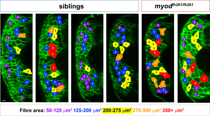
Nuclear counts and size measurement of fast fibres. Transverse views of wholemount 5 dpf larvae from an incross of myodfh261/+;Tg(Ola.Actb:Hsa.HRAS-EGFP)vu119 injected at 1 cell stage with RNA encoding H2B-mCherry (similar to Addgene #20972, red channel not shown). At 5 dpf, fully dual-labelled larvae were scanned in 3D confocal stacks and retrospectively genotyped. Coloured fibres indicate those selected for measurement using the criteria of a) unambiguously delineation of plasma membrane and nuclei and b) distribution throughout the fast myotome of somite 17. The cross-sectional area at the anteroposterior midpoint of each fibre was measured using Volocity v6.3 and the number of nuclei counted manually in twenty fibres in each fish. Note that fibre outline does not perfectly match the shape of fibre because the midpoints of most fibres are in different planes. Size of fibre is colour coded and nuclear number indicated on each fibre. Data for all sibling (80) and all mutant (40) fibres were pooled and presented in Fig. 6F-H. Scale bar = 10 µm.
Reprinted from Developmental Biology, 431(2), Roy, S.D., Williams, V.C., Pipalia, T.G., Li, K., Hammond, C.L., Knappe, S., Knight, R.D., Hughes, S.M., Myotome adaptability confers developmental robustness to somitic myogenesis in response to fibre number alteration, 321-335, Copyright (2017) with permission from Elsevier. Full text @ Dev. Biol.