
Image
|
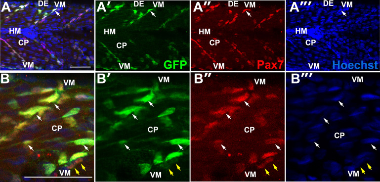
Figure Caption
Fig. s4
pax7a:GFP+ cells co-express Pax7. Wholemount 3.25 dpf pax7a:GFP;pfe/pfe larvae in lateral view with dorsal up and anterior to left after immunodetection of GFP and Pax7, and counterstaining with Hoechst 33342. Single deep confocal slices of two whole somites (A) and epaxial somite (B) are shown. GFP largely co-localizes with Pax7 (white arrows). Pax7+ cell on VM that is not GFP+ (yellow arrows). Scale bars = 50 µm.
Acknowledgments
This image is the copyrighted work of the attributed author or publisher, and
ZFIN has permission only to display this image to its users.
Additional permissions should be obtained from the applicable author or publisher of the image.
Reprinted from Developmental Biology, 431(2), Roy, S.D., Williams, V.C., Pipalia, T.G., Li, K., Hammond, C.L., Knappe, S., Knight, R.D., Hughes, S.M., Myotome adaptability confers developmental robustness to somitic myogenesis in response to fibre number alteration, 321-335, Copyright (2017) with permission from Elsevier. Full text @ Dev. Biol.