
|
Fig. 7
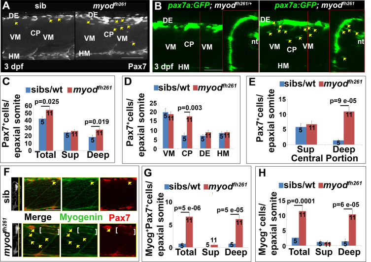
Premature ingression of Pax7+ myogenic precursors in myodfh261 mutants. Wholemount larvae from a myodfh261/+ incross stained for Pax7 (A,C-H) or pax7a:GFP;myodfh261/+ incross imaged live (B) at 3 dpf and shown in confocal short stacks in lateral view. A. Pax7 antibody stained nuclei (arrows) within the deep CP myotome. B. Pax7:GFP+ cells (arrows) in lateral and transversal views. C-H. Comparison of Pax7+ (C-E), Myog+Pax7+ (G) and Myog+ (H) cell numbers in the epaxial half of som16-18 between the number of myodfh261 mutants and their siblings indicated within columns. The extra Pax7+ cells in myodfh261 mutants (C) were specifically located in deep CP (D,E). Lateral planes at deep locations indicated by yellow line on transversal sections of wholemount larvae stained for Pax7, Myogenin and Hoechst (F) reveal increased numbers of Myog+ and Pax7+Myog+ in the deep CP (arrows). Note the alignment of some Myog+ nuclei (brackets). VM: vertical myosepta, CP: central portion, DE: dorsal edge, HM: horizontal myoseptum. Bars 50 µm.
Reprinted from Developmental Biology, 431(2), Roy, S.D., Williams, V.C., Pipalia, T.G., Li, K., Hammond, C.L., Knappe, S., Knight, R.D., Hughes, S.M., Myotome adaptability confers developmental robustness to somitic myogenesis in response to fibre number alteration, 321-335, Copyright (2017) with permission from Elsevier. Full text @ Dev. Biol.