
|
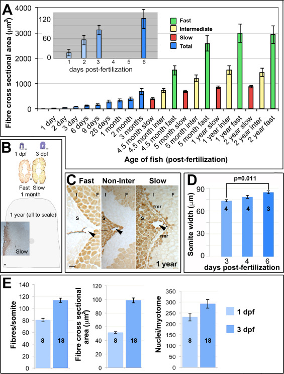
Fig. 1
Muscle growth involves increase in fibre number and size. A. Fibre cross sectional area from unfixed cryosections as a function of age and fibre type in single average-sized fish at each age. Mean±SEM from 80 to 250 fast fibres and all slow and intermediate fibres in midbody somites. Inset magnifies early stages. B. Toluidine blue and myosin stained midbody sections at same scale Schematic indicates approximate size of entire section at 1 year. C. Immunodetection distinguishes fast (F), intermediate (I) and slow (S) fibres including the red muscle rim (rmr) from 4.5 months. Arrowheads indicate the lateral line. D. Mediolateral width of somite measured at horizontal myoseptum from wholemount confocal stacks. E. Fibres and nuclei were counted and cross sectional area measured on YZ confocal sections of somite 16–20 from 8 and 18 lightly-fixed Hoechst-stained Tg(Ola.Actb:Hsa.HRAS-EGFP) embryos at 1 and 3 dpf, respectively. As small fibres are hard to count with confidence in fixed preparations, fibre numbers represent minimal estimates. Note that the 1 dpf values for fibre cross sectional area are significantly larger in wholemount measurements (E) compared to cryosectioned material (A), possibly due to methodological and/or lay-to-lay variation. Bars 50 µm.
Reprinted from Developmental Biology, 431(2), Roy, S.D., Williams, V.C., Pipalia, T.G., Li, K., Hammond, C.L., Knappe, S., Knight, R.D., Hughes, S.M., Myotome adaptability confers developmental robustness to somitic myogenesis in response to fibre number alteration, 321-335, Copyright (2017) with permission from Elsevier. Full text @ Dev. Biol.