
Image
|
Figure Caption
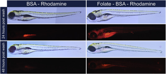
Fig. 5
Folate targeted compounds are selectively taken up and maintained in the embryo. Brightfield and fluorescence representative images of wild-type fish exposed to either BSA conjugated to rhodamine dye or folate conjugated to BSA and rhodamine dye. After 24 h exposure to BSA based compounds, embryos were washed and transferred to standard embryo media. Exposed embryos were then imaged at 24 and 48 h after being transferred to standard embryo media and shown above.
Acknowledgments
This image is the copyrighted work of the attributed author or publisher, and
ZFIN has permission only to display this image to its users.
Additional permissions should be obtained from the applicable author or publisher of the image.
Reprinted from Gene expression patterns : GEP, 25-26, Jones, R.N., Erhard, S.A., Malham, M.R., Gen, A.Y., Sullivan, K., Olsen, K.W., Dale, R.M., Expression and characterization of the zebrafish orthologue of the human FOLR1 gene during embryogenesis, 159-166, Copyright (2017) with permission from Elsevier. Full text @ Gene Expr. Patterns