
Image
|
Figure Caption
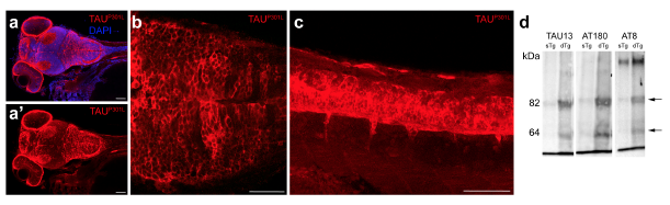
Fig. s12 (a) Immunohistochemical staining for TAUP301L in 9 day-old dTg larvae. Anterior view of the brain is shown. (b) Single fluorescence channel for TAUP301L. (c) High-magnification image of the frame in b. (d) TAUP301L expression in the spinal cord. (e) Western blot analyses of TAUP301L (TAU13), and hyperphosphorylated form of TAU with AT180 and AT8. Scale bars equal 50 μm.
Acknowledgments
This image is the copyrighted work of the attributed author or publisher, and
ZFIN has permission only to display this image to its users.
Additional permissions should be obtained from the applicable author or publisher of the image.
Full text @ Sci. Rep.