
|
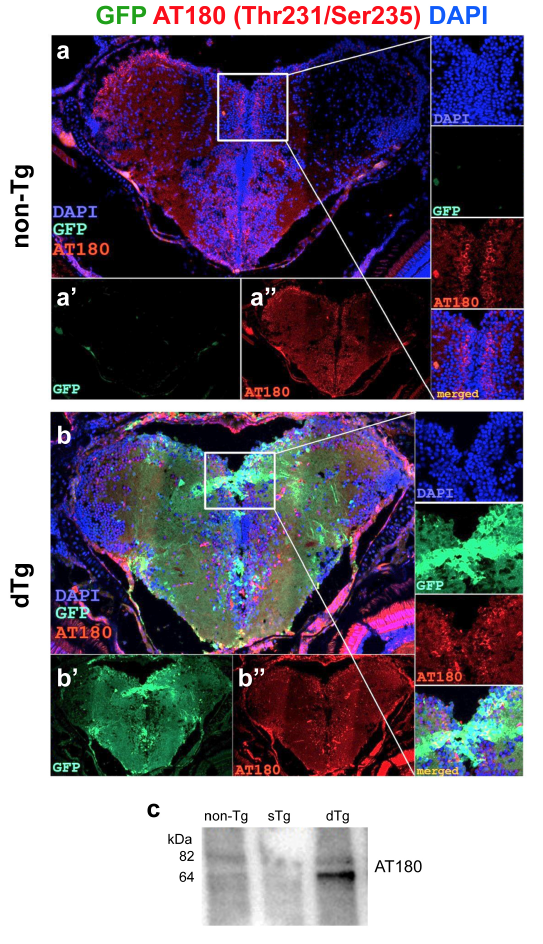
Fig. s7 (a) Immunohistochemistry (IHC) for AT180 (red) and GFP (green) on coronal sections of telencephalon of a 6-month old nontransgenic animal. (a’, a’’) Individual fluorescent channels for GFP (a’) and AT180 (a’’). Insets show the enlarged view of the frame in A with individual channels for DAPI, GFP and AT180, and merged image. (b) IHC for AT180 and GFP on coronal sections of telencephalon of a 6-month old dTg animal. (b’, b’’) Individual fluorescent channels for GFP (b’) and AT180 (b’’). Insets show the enlarged view of the frame in b with individual channels for DAPI, GFP and AT180, and merged image. (c) Western blot for AT180 from brains of non-Tg, sTg and dTg animals. Scale bars equal 25 μm. n = 5 fish for every staining.