
Image
|
Figure Caption
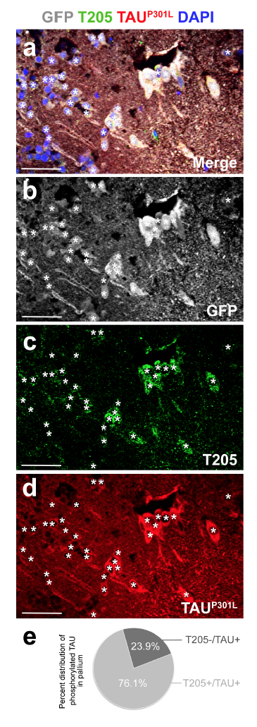
Fig. s6 Immunohistochemical staining for GFP (recombined cells, white), T205 (hyperphosphorylated TAU, green), and TAUP301L (red) in 6 month-old dTg animal. (a) merge image. Individual channels for (b) GFP, (c) T205, and (d) TAUP301L. (e) Quantification of the percentage of TAUP301Lpositive cells that are T205 positive or negative. Scale bars equal 50 μm. n = 3 fish and >20 histological sections for every staining and quantification.
Acknowledgments
This image is the copyrighted work of the attributed author or publisher, and
ZFIN has permission only to display this image to its users.
Additional permissions should be obtained from the applicable author or publisher of the image.
Full text @ Sci. Rep.