
|
Fig. 4
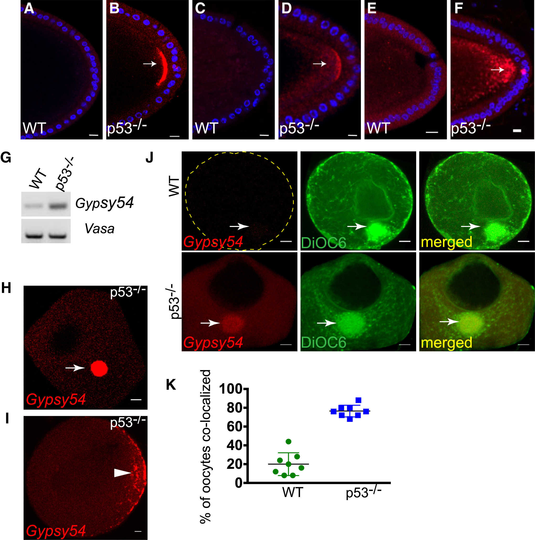
Germ Plasm Localization Is Conserved in Invertebrates and Vertebrates
(A–F) Patterns of other retrotransposons in the fly oocyte were profiled by in situ hybridization. Examples of Tahre (A and B), Burdock (C and D), and HeT-A (E and F) on WT (A, C, and E) and p53−/− (Β, D, and F) oocytes are shown. DAPI counterstain is indicated in blue. Like Tahre, Burdock, and HeT-A RNAs also localized to the germ plasm region (arrows).
(G–K) Gypsy54 localizes to the germ plasm of zebrafish oocytes. (G) RT-PCR analysis of Gypsy54 and vasa (loading control) from WT and p53−/− zebrafish ovaries. (H)–(J) show in situ hybridizations for Gypsy54 RNA (red) on WT and p53−/− oocytes. Arrows point to the Balbiani body, the presumptive germ plasm of stage I oocytes (H). The arrowhead in (I) indicates the germ plasm at the cortical region of stage II oocytes. DiOC6 (green) was used to label the Balbiani body (J, arrows) in p53−/− and WT oocytes. Images are single optical sections. The scatterplot in (K) shows percentages of DiOC6-stained Balbiani bodies that were also positive for Gypsy54 RNA.
Scale bars, 10 μm. See also Figure S4 and STAR Methods.