
Image
|
Figure Caption
Fig. 5
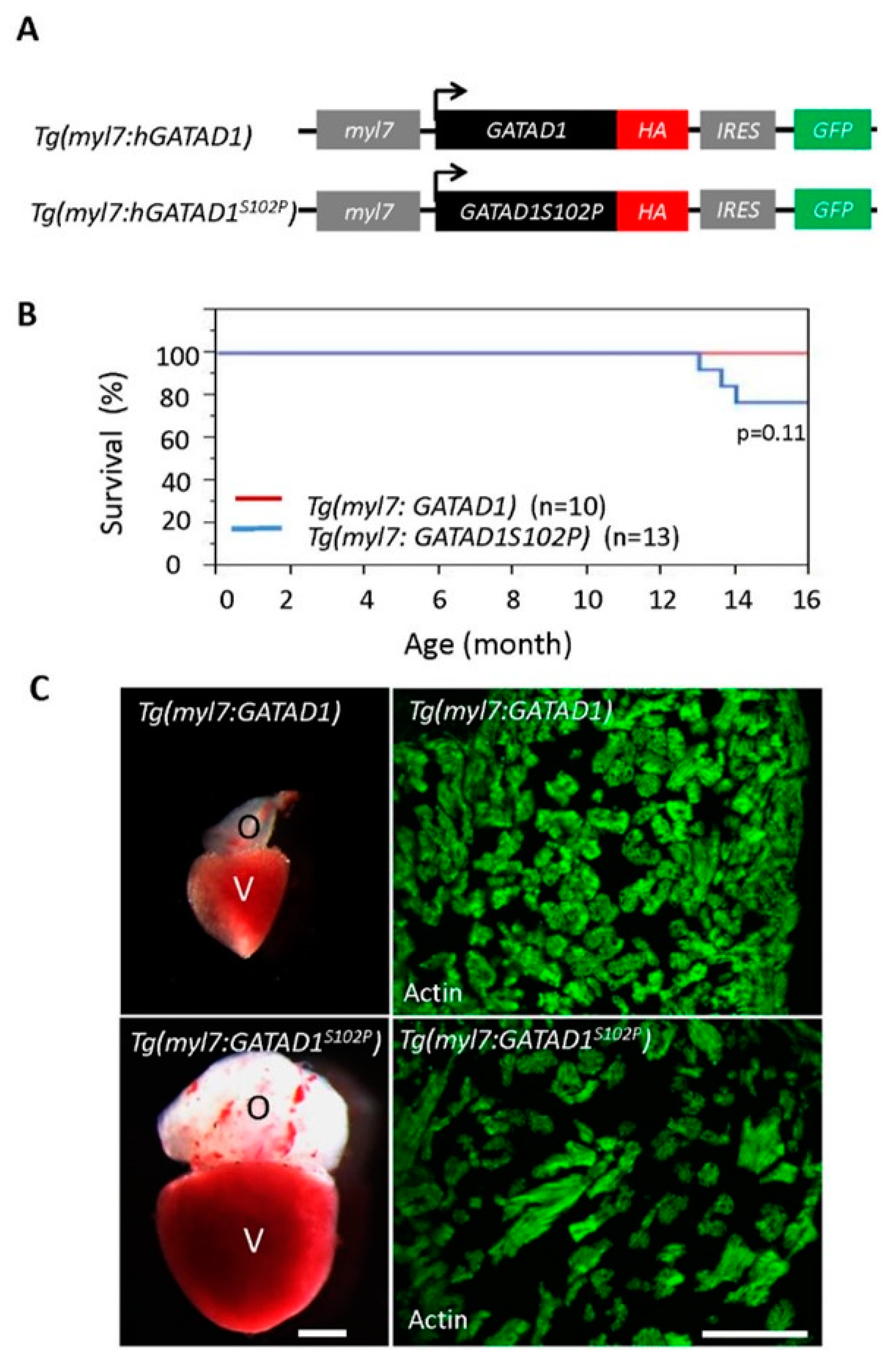
Generating and phenotyping GATAD1 transgenic fish. (A) Schematic illustration of constructs that were used to generate two transgenic fish lines expressing either human wild type GATAD1 or GATAD1-S102P mutation. The GATAD1 gene is flanked by the myl7 enhancer at its 5′ terminal to drive cardiomyocyte-specific expression, and it has an HA tag and IRES-EGFP at its 3′ terminal to facilitate detection of ectopic gene expression and fish propagation; (B) The transgenic fish expressing mutant GATAD1 started to die at approximately 12 months of age, and all transgenic fish expressing wild type GATAD1 were able to survive to 16 months; (C) Severe cardiac hypertrophy in a Tg(myl7:hGATAD1S102P) fish. Left panel, images of a heart dissected from a Tg(myl7:hGATAD) and a Tg(myl7:hGATAD1S102P) fish at 17 months of age, separately. The heart from this single Tg(myl7:hGATAD1S102P) fish exhibits significantly enlarged ventricle and out flow tract. Right panel, phalloidin staining revealed less dense myofibril and wider myofibril in the heart of this single Tg(myl7:hGATAD1S102P) fish. V, ventricle; O, out flow tract; Scale bar 0.5 mm.
Figure Data
Acknowledgments
This image is the copyrighted work of the attributed author or publisher, and
ZFIN has permission only to display this image to its users.
Additional permissions should be obtained from the applicable author or publisher of the image.
Full text @ J Cardiovasc Dev Dis