
Image
|
Figure Caption
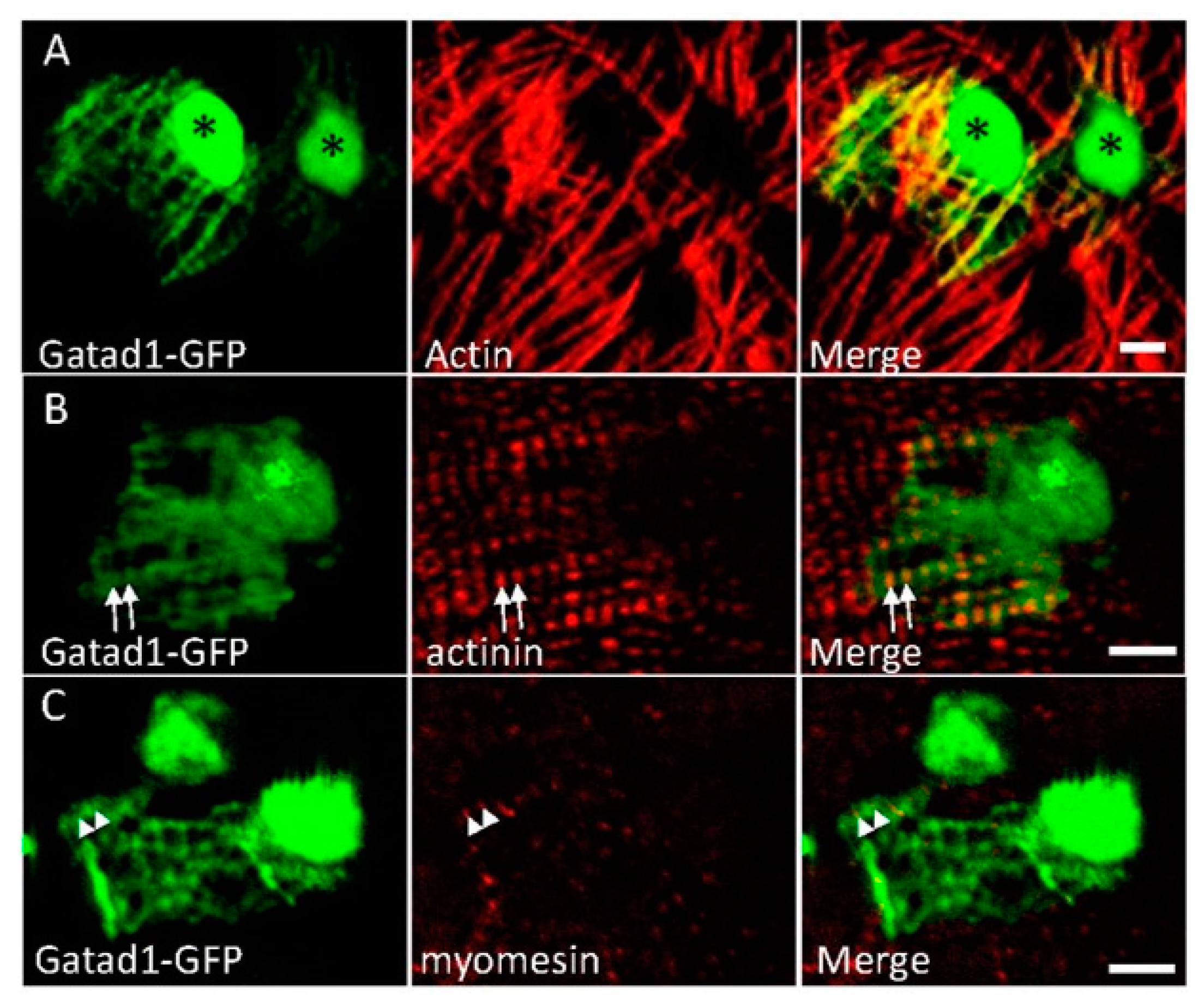
Fig. 3
Subcellular localization of the Gatad1 protein in the embryonic zebrafish heart. After the myl7:gatad1-GFP construct was injected into 1-cell staged embryos, hearts from 2 dpf embryos were dissected for immunostaining and imaging. (A) Gatad1-GFP shows strong expression in nuclei and relatively weak expression in myofibrils, overlapping with Actin as revealed by phalloidin staining; (B) Gatad1-GFP partially overlaps with Z-discs marked by Actinin (arrows); (C) Gatad1-GFP forms alternatively striated patterns with M-line marked by Myomesin (arrowheads). * Nucleus; Scale bar 5 μm.
Acknowledgments
This image is the copyrighted work of the attributed author or publisher, and
ZFIN has permission only to display this image to its users.
Additional permissions should be obtained from the applicable author or publisher of the image.
Full text @ J Cardiovasc Dev Dis