
|
Fig. S3
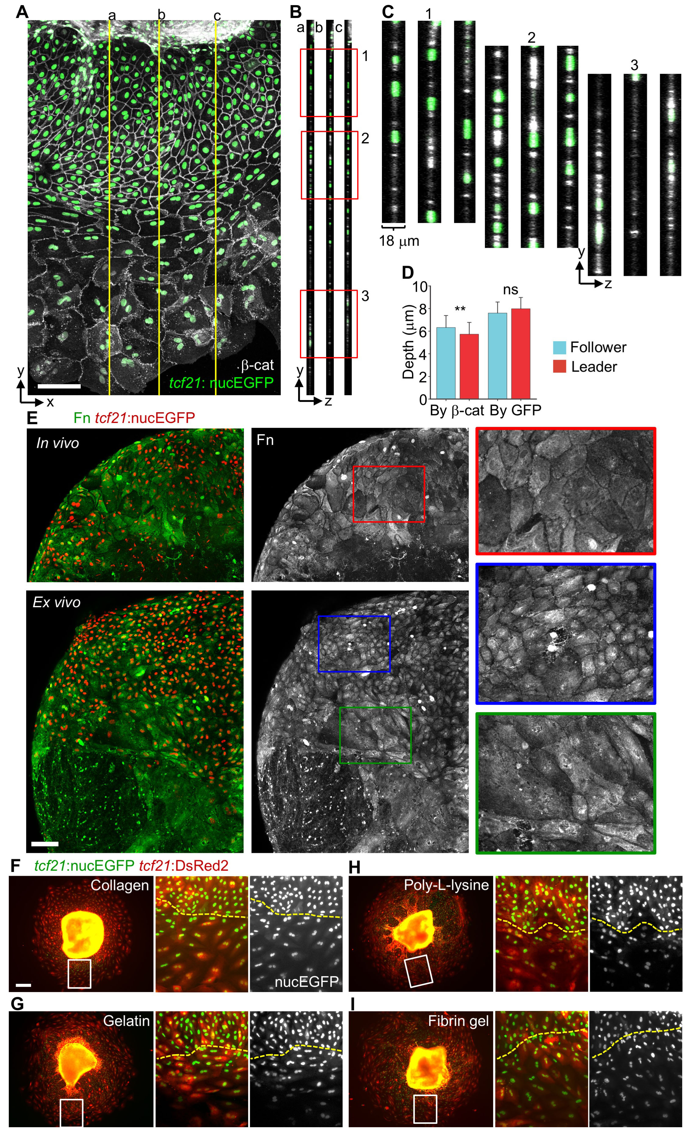
Analysis of Cell Volume and the Relevance of ECM Components to Epicardial Regeneration (Related to Figure 2)
(A) A maximum projection view of an explant culture, showing tcf21:nucEGFP in green and b-catenin staining in grayscale. Three yellow lines (a, b and c) denote positions of views for the y-z planes in (B).
(B, C) y-z views for the planes denoted in (A). The framed regions in (B) are enlarged to show details in (C).
(D) Quantification of the cell depths by measuring junctions (b-catenin signals, By b-cat) or nuclei (nucEGFP signals, By GFP). n = 50 (By b-cat, follower), 64 (By b-cat, leader), 25 (By GFP, follower) and 10 (By GFP, leader), respectively. ** P < 0.01; ns, not significant; Mann-Whitney Rank Sum Test. Bars indicate mean ± S.D.
(E) Images of fibronectin (Fn) staining of regenerating hearts both in vivo and ex vivo. Fn is shown in green (merged images) or grayscale (single-channel images) and tcf21:nucEGFP is shown in red. The framed regions are enlarged to show details on the right. Scale bar, 100 μm.
(F-I) tcf21:nucEGFP; tcf21:DsRed2 epicardial explants were plated in differently coated dishes for 5 days. Dishes were coated with 0.01% type I collagen (F), 0.1% gelatin (G) or 0.01% poly-L-lysine (H) solutions overnight. Fibrin gel (I) was freshly made as described previously (Kim, et al., 2012). The framed regions of left panels are enlarged to show as middle (merged) and right panels (nucEGFP only). The yellow lines approximately separate follower (top) and leader cell regions (below). Scale bar, 100 μm.
Reprinted from Developmental Cell, 42, Cao, J., Wang, J., Jackman, C.P., Cox, A.H., Trembley, M.A., Balowski, J.J., Cox, B.D., De Simone, A., Dickson, A.L., Di Talia, S., Small, E.M., Kiehart, D.P., Bursac, N., Poss, K.D., Tension Creates an Endoreplication Wavefront that Leads Regeneration of Epicardial Tissue, 600-615.e4, Copyright (2017) with permission from Elsevier. Full text @ Dev. Cell