
|
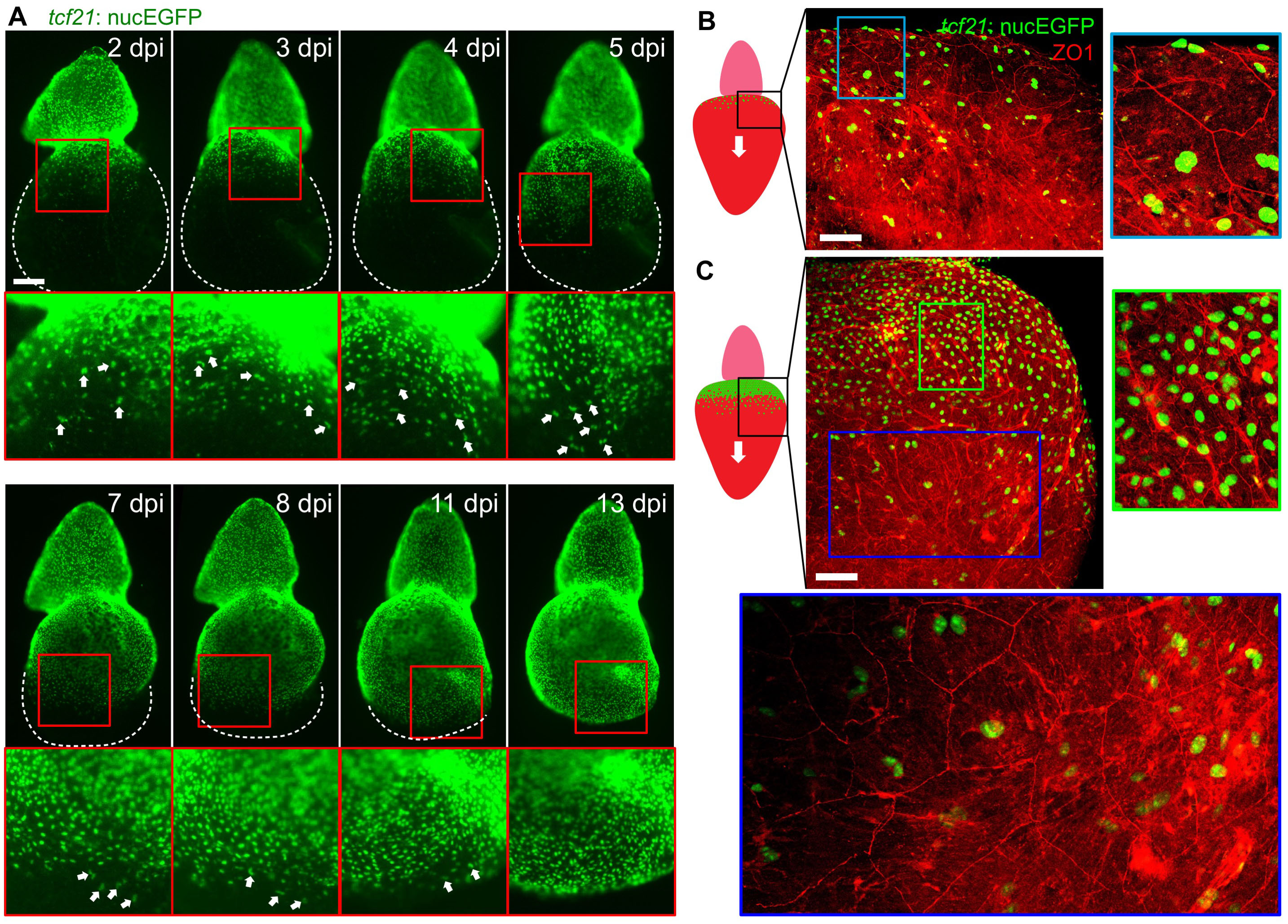
Fig. S2
Cell Size and Nuclear Number during ex vivo Epicardial Regeneration (Related to Figure 1)
(A) A whole ventricular explant (with outflow tract at top) undergoing ex vivo epicardial regeneration. tcf21:NTR; tcf21:nucEGFP hearts were incubated in culture medium with 1 mM Mtz for 24 h. Explants were assessed daily for EGFP fluorescence. White dashed lines outline ventricles. Regions framed in red are enlarged to show details below each panel. White arrows denote nuclear doublets. Scale bar, 200 μm.
(B, C) Images of flattened explants stained with an anti-ZO1 antibody (red). Images of the framed region represented in the cartoons from 2 hearts are shown. Regions framed in cyan, green and blue were enlarged in the same scale at the right or the bottom. Optical sections are shown in enlarged images in (C) to more clearly indicate ZO1 staining. Scale bar, 100 μm.
Reprinted from Developmental Cell, 42, Cao, J., Wang, J., Jackman, C.P., Cox, A.H., Trembley, M.A., Balowski, J.J., Cox, B.D., De Simone, A., Dickson, A.L., Di Talia, S., Small, E.M., Kiehart, D.P., Bursac, N., Poss, K.D., Tension Creates an Endoreplication Wavefront that Leads Regeneration of Epicardial Tissue, 600-615.e4, Copyright (2017) with permission from Elsevier. Full text @ Dev. Cell