
|
Fig. 6 Follower Cells Undergo Endoreplication after Leader Cell Ablation
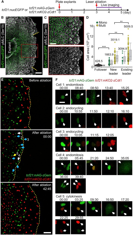
(A) Schematic for experiments in (B) to (F).
(B and C) Ablation experiment using tcf21:nucEGFP explants (green). Twenty-four hours post laser ablation, the explant culture was stained to detect pMLC (Ser19, grayscale). The framed regions in (B) are enlarged to show details in (C). The yellow dashed lines approximately mark the region that was ablated. Scale bars, 200 μm (B) and 100 μm (C).
(D) Quantification of nucleation and cell area of de novo leader cells and pre-existing leader and follower cells in (B). Leader cell region is defined by the strong staining of pMLC. Mono, mononucleate; Multi, multinucleate. n = 111 (follower), 39 (new leader, Mono), 49 (new leader, Multi), 10 (existing leader, Mono), and 89 (existing leader, Multi), respectively. ∗∗∗p < 0.001; ∗∗p < 0.01; Mann-Whitney rank-sum test. Error bars indicate SD.
(E) Video frames of a tcf21:FUCCI epicardial explant culture subjected to live imaging and laser ablation. The top panel is an image before ablation; the middle panel, immediately after ablation; the lower panel, a reconstructed leader cell region at 42 hr 45 min after ablation. The white, cyan, and yellow arrows indicate nuclei that underwent endomitosis, endocycling, and cytokinesis, respectively. Timing, hr:min. Scale bar, 100 μm.
(F) Cropped video frames of the numbered cells indicated in the middle panel of (E). White arrows indicate the nuclei of cells that undergo endoreplication or cytokinesis. Timing, hr:min. Scale bar, 20 μm.
See also Movie S7.
Reprinted from Developmental Cell, 42, Cao, J., Wang, J., Jackman, C.P., Cox, A.H., Trembley, M.A., Balowski, J.J., Cox, B.D., De Simone, A., Dickson, A.L., Di Talia, S., Small, E.M., Kiehart, D.P., Bursac, N., Poss, K.D., Tension Creates an Endoreplication Wavefront that Leads Regeneration of Epicardial Tissue, 600-615.e4, Copyright (2017) with permission from Elsevier. Full text @ Dev. Cell