
|
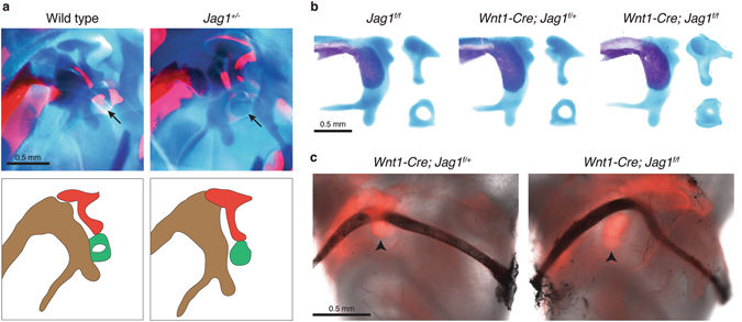
Fig. 2 Mispatterning of middle ear cartilages and formation of the stapedial artery in Jag1-deficient mice. (a,b) Newborn mice were stained with Alcian Blue for cartilage and Alizarin Red S for bone. Close-up views show the developing middle ear, which is diagrammed below for wild-type and Jag1 heterozygous mice (malleus, brown; incus, red; stapes, green). Dissected middle ear cartilages are shown for conditional mutants. Arrows point to the stapes cartilage, which is reduced in size in both heterozygous and conditional Jag1 mutant mice. (c) The stapes of Wnt1-Cre; Jag1 f/+; Rosa26-Tomato and Wnt1-Cre; Jag1 f/f; Rosa26-Tomato mice fluoresce red and the stapedial arteries appear black from India ink injection. The artery is still present in Jag1-CKO mice, where it deviates around the misshapen stapes cartilage. Arrowheads point to the stapes.