
|
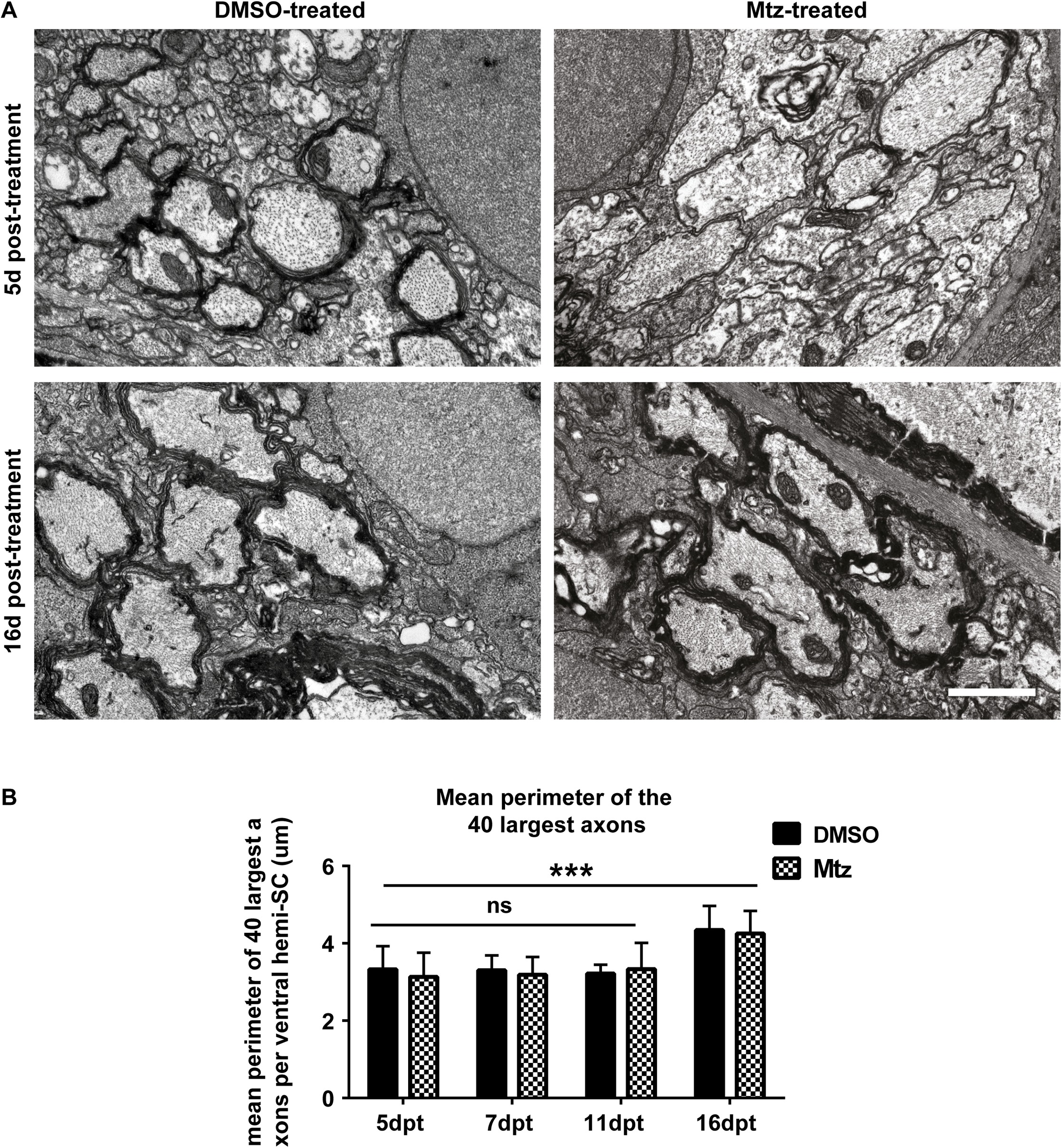
Fig. 9
Remyelination coincides with growth of axon caliber between 5dpt and 16dpt.
A. Top panel: representative electron micrographs from DMSO- and Mtz-treated Tg(mbp:mCherry-NTR) larvae at 5dpt. Bottom panel: same from larvae 16dpt. Scale bar: 1μm. B. Quantification of the mean perimeter of the 40 largest axons (whether myelinated or unmyelinated) in control and treated larvae at 5, 7, 11 and 16dpt. At 5dpt, mean perimeter of the 40 largest axons in controls: 3.32 μm ± 0.60 μm and treated 3.13 μm ± 0.62 μm. At 7dpt, controls 3.30μm ± 0.39μm and treated 3.19μm ± 0.46μm. At 11dpt, controls 3.21μm ± 0.23μm and treated 3.33μm ± 0.68μm. At 16dpt, the mean perimeter of the largest 40 axons in controls was 4.34 ± 0.63 in controls and in treated, 4.25 ± 0.59. A two-way ANOVA found a significant main effect of time point (p = 0.0003) but a non-significant main effect of treatment condition (p = 0.707) and a non-significant interaction (p = 0.952).