
|
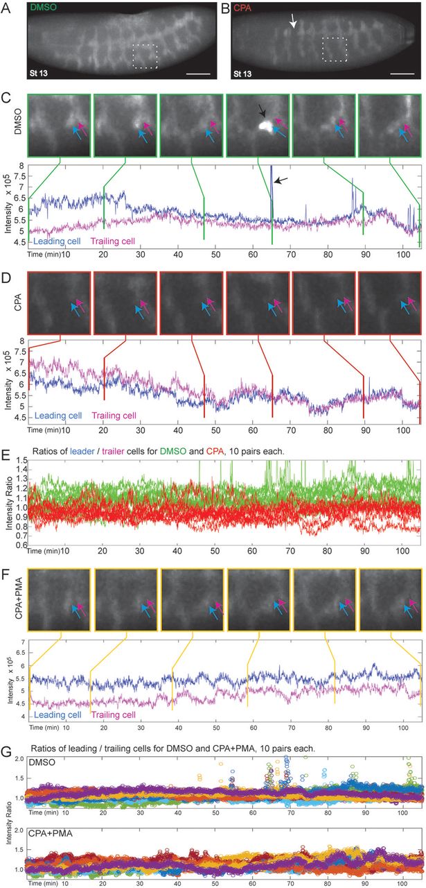
Fig. 3 SERCA regulates cell migration and budding by maintaining higher Ca2+ levels in ‘leader’ versus ‘trailer’ cells. (A,B) Btl:GCamp3 embryos treated with DMSO, CPA, or CPA+PMA were imaged between stage 13 and 16, and images were reconstructed to analyze the Ca2+ levels in tracheal cells. Shown are 3D reconstructed images of stage 13 embryos treated with (A) DMSO or (B) CPA (arrow indicates discontinuous trunk). Insets mark segments tracked in (C) and (D). Scale bars: 50 µm. (C-D,F) Inset images of tracked cells from DMSO- (C), CPA- (D), and CPA+PMA-treated (F) embryos. ‘Leader’ cells (blue arrow) that migrate from adjacent segments and fuse to form the lateral trunk and ‘trailer’ cells (magenta arrow) behind them were tracked over the time course and their Ca2+ levels quantified and plotted below the panel of images. Vertical lines mark the time points corresponding to each image. (C) In controls, there is a Ca2+ level differential whereby ‘leader’ cells have consistently higher Ca2+ levels than ‘trailers’, particularly early when the cells are migrating. After fusion, ‘leaders’ periodically display surges of Ca2+ (black arrows; also green spikes in E and spikes in top panel of G). (D) In CPA-treated embryos, migration is lost so the lateral trunk remains discontinuous, and ‘leaders’ have lower Ca2+ levels than ‘trailers’ during the time they should be migrating. Thus the Ca2+ level differential is reversed. (F) CPA+PMA co-treatment reinstates the higher Ca2+ level in ‘leaders’ and rescues migration. (E,G) For each treatment, the ratio of intensities of ten pairs of cells (leader/trailer) was plotted. (E) The DMSO ratios (green) average >1. SERCA inhibition (red, ratio <1) inverts this. (G) The DMSO- and CPA+PMA-treated embryos overlap (ratios >1), so each was plotted separately with different colors for individual ratios. DMSO-treated embryos show Ca2+ spikes following trunk fusion. While CPA+PMA restores branching, Ca2+ spikes are absent or only seen much later (Movie 5).