
Image
|
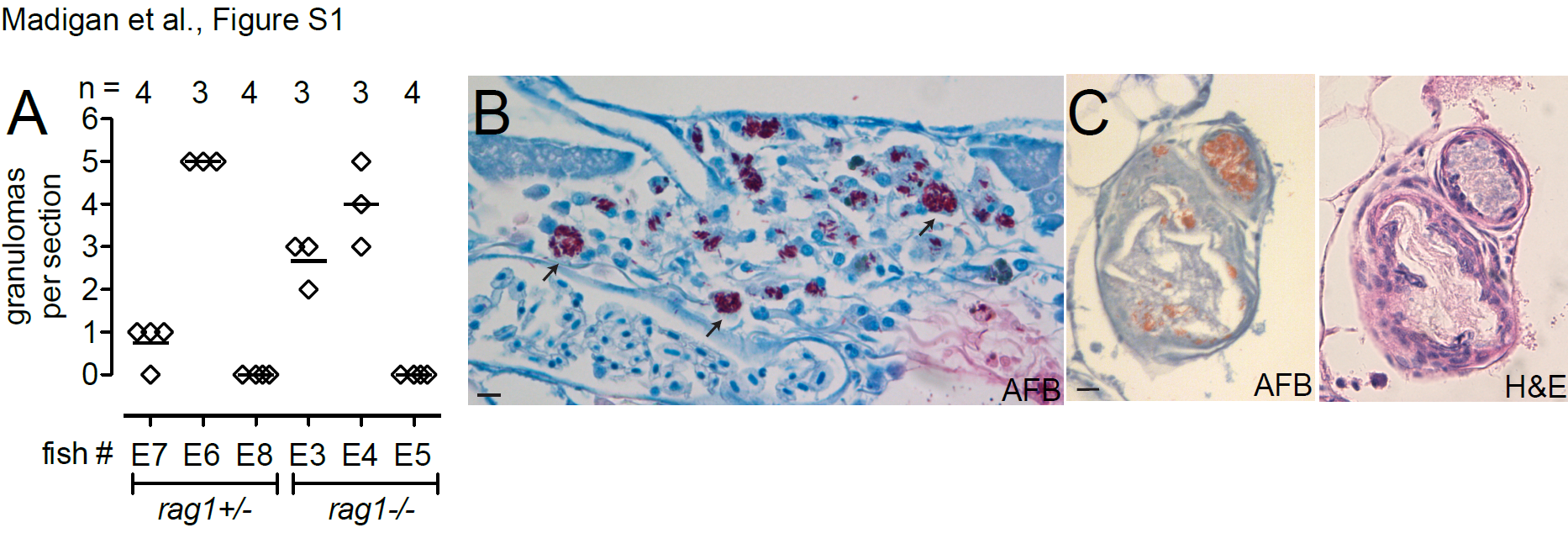
Figure Caption
Fig. S1
Detailed analysis of M. leprae granulomas. In panel A, multiple AFB-stained sections from infected fish at 112 dpi were scored for number of infected granulomas; n, number of sections scored. In panel B, an AFB-stained section of a non-necrotizing granuloma in a rag1 heterozygote zebrafish with heavily infected macrophages (arrows). In panel C, AFB and H&E sections of a necrotic granuloma observed in M. leprae-infected rag1 heterozygote fish. 10μm bars.
Acknowledgments
This image is the copyrighted work of the attributed author or publisher, and
ZFIN has permission only to display this image to its users.
Additional permissions should be obtained from the applicable author or publisher of the image.
Full text @ J. Infect. Dis.