
|
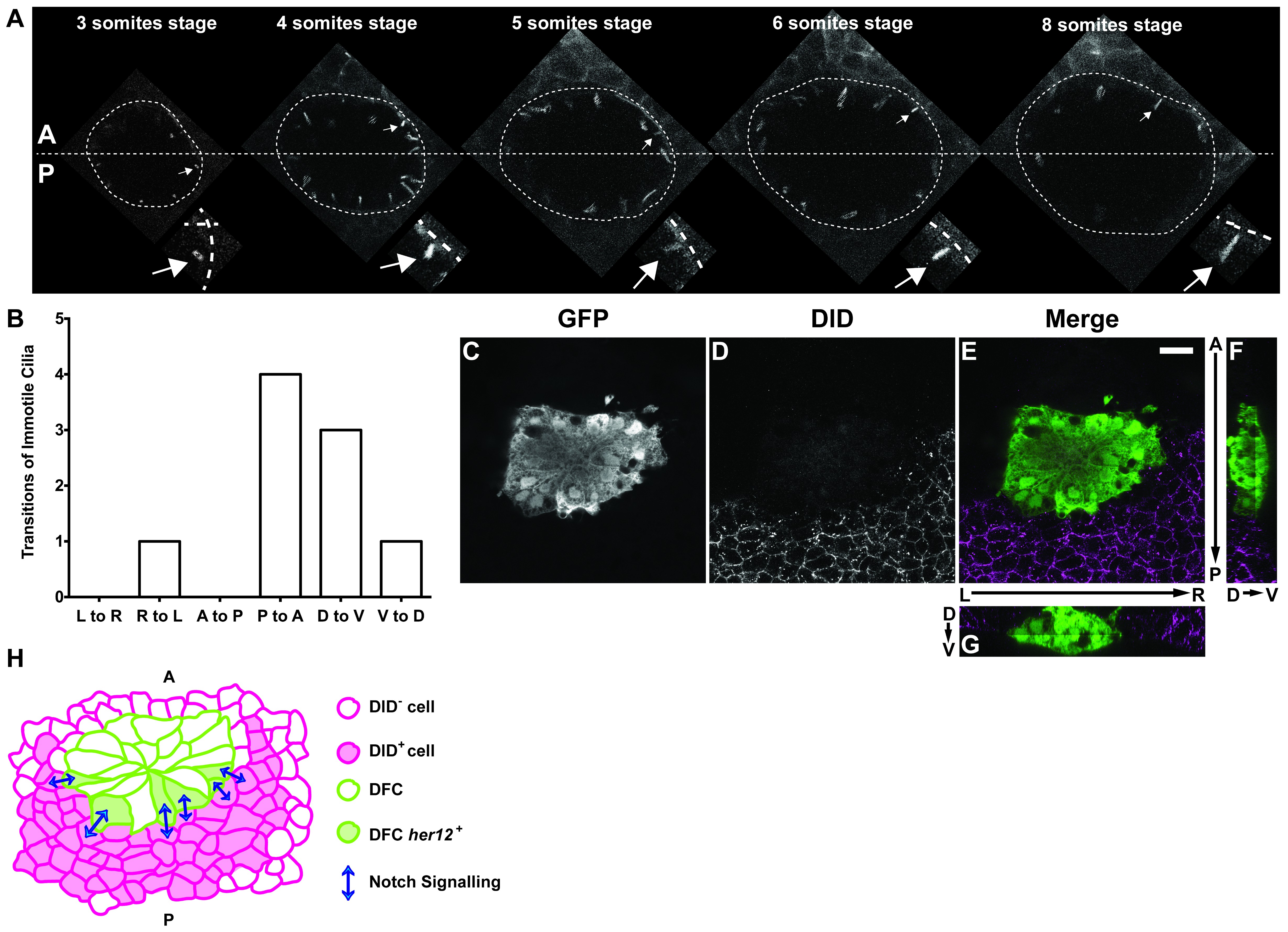
Fig. 5 S2
Positions of immotile cilia in the anterior-posterior axis change through development.
(A) Transition of an immotile cilium from a Posterior position to an ever more Anterior position as development of the embryo progresses from 3 ss to 8 ss. (B) Number and type of transitions of Immotile cilia along the three axes (Anterior – Posterior; Dorsal – Ventral; Left – Right) from 3 to 8 somites stage (4 embryos, 9 cilia transitions, 19 immotile cilia tracked). L to R – Left to Right; R to L – Right to Left; A to P – Anterior to Posterior; P to A – Posterior to Anterior; D to V – Dorsal to Ventral; V to D – Ventral to Dorsal. (C–G) Localization of the DlD ligand by immune-histochemistry with an antibody anti-DlD (D) in sox17:GFP transgenic embryos at bud stage in Control. An antibody anti-GFP was simultaneously used in order to highlight the DFCs (C), with the resulting merged image in (E). Scale bar represents 20 μm. Anterior is to the top and Left is to left. (F–G) Orthogonal projections emphasising DLD expression. (F) Anterior is to the top and Dorsal is to left. (G) Dorsal is to the top and Left is to left. (H) Model depicting how the NS is occurring between the DLD positive surrounding cells and the Notch positive DFC in the posterior region of the cluster.