
Image
|
Figure Caption
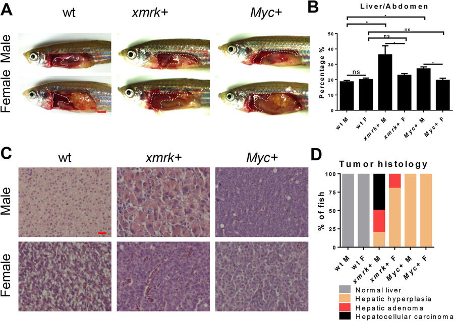
Fig. 1
Characterization of sex disparity in xmrk and Myc-induced HCC progression. Three-month-old, xmrk+, Myc+ and wildtype zebrafish were treated with 60 μg/ml dox for 7 days. 10 fish were analysed in each group and the experiment was repeated multiple times. (A) Gross morphology of male and female fish (left lateral view). The livers are outlined. (B) Quantification of percentage of liver area to abdomen area. (C) Representative images of liver sections of male and female fish after H&E staining. (D) Quantification of tumor histology in male and female fish. *P < 0.05. Scale bars: 2 mm in (A) and 20 μm in (C).
Acknowledgments
This image is the copyrighted work of the attributed author or publisher, and
ZFIN has permission only to display this image to its users.
Additional permissions should be obtained from the applicable author or publisher of the image.
Full text @ Sci. Rep.